






For individuals navigating the often-turbulent waters of hypermobile Ehlers-Danlos syndrome (hEDS) and related hypermobility spectrum disorders (HSD), the body frequently sends complex, sometimes alarming, signals. Among the most common visual concerns is the sudden, striking redness of the hands and feet. While many patients are familiar with the phenomenon of "blood pooling"—a byproduct of the dysautonomia that frequently accompanies hypermobility—a rarer and more painful condition known as erythromelalgia often lurks in the background, frequently misdiagnosed or overlooked.
As our understanding of the autonomic nervous system evolves, so too does the recognition that erythromelalgia is not merely a cosmetic concern, but a significant neuropathic condition that shares a deep, biological kinship with the hEDS community.
Main Facts: Distinguishing Pain from Pooling
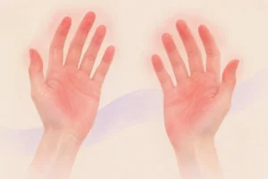
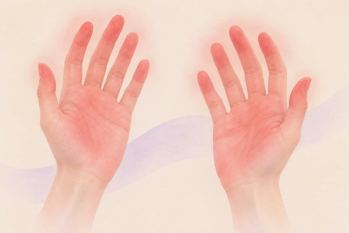
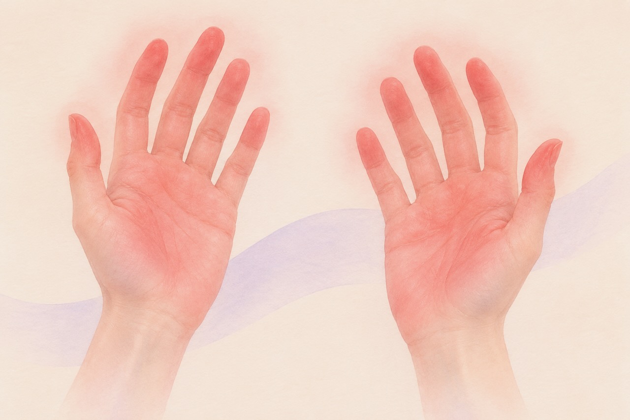
The clinical challenge lies in the visual similarity between blood pooling and erythromelalgia. Both manifest as a reddening of the extremities, but their underlying mechanisms are distinct.
Blood pooling is a common manifestation of dysautonomia, where the autonomic nervous system fails to adequately constrict blood vessels or support venous return. When an individual stands or lets their limbs hang, gravity causes blood to settle in the extremities, resulting in a pink, purple, or mottled discoloration. This is typically painless or associated with only mild discomfort and, crucially, resolves almost immediately upon elevation or lying down.
Erythromelalgia (EM), by contrast, is a distinct neurovascular disorder characterized by the "triad" of intense redness, localized warmth, and severe burning pain. Unlike the passive settling of blood in venous insufficiency, EM involves a dysfunctional nervous response. It is frequently triggered by environmental or internal heat, exercise, emotional stress, or even the consumption of stimulants like caffeine and alcohol. The burning sensation is often described as searing, stabbing, or electrical, and unlike blood pooling, it does not reliably dissipate with simple elevation.
Chronology of Clinical Understanding
The history of erythromelalgia is a journey from anecdotal observation to modern neurological classification.
- Late 19th Century: The condition was first described by Silas Weir Mitchell in 1878, who coined the term "erythromelalgia" (from the Greek erythros meaning red, melos meaning limb, and algos meaning pain).
- Mid-20th Century: Researchers began to differentiate between primary (inherited or idiopathic) and secondary (associated with underlying systemic disease) forms of the disorder.
- The Genomic Breakthrough (2004): A landmark discovery identified mutations in the SCN9A gene in families with primary inherited erythromelalgia. This revealed that the condition was fundamentally a channelopathy—a disorder of the voltage-gated sodium channels responsible for transmitting pain signals.
- The Modern Era (2020–2026): Recent clinical research has bridged the gap between EM and the hypermobility community. Studies, including a pivotal 2025 analysis of skin biopsies, have confirmed that EM patients frequently exhibit the same small fiber loss seen in small fiber neuropathy (SFN) and POTS, cementing its status as a recognized dysautonomia syndrome.
Supporting Data: The Biological Link to hEDS
The connection between hEDS, HSD, and erythromelalgia is rooted in the shared vulnerability of the nervous system. Small fiber neuropathy (SFN)—a condition where the small-diameter, unmyelinated nerve fibers responsible for pain, temperature, and autonomic control are damaged—is highly prevalent in the hEDS population.
The Anatomy of an Episode
Research indicates that in secondary erythromelalgia, the damage to these small nerve fibers disrupts the body’s ability to regulate the constriction and dilation of blood vessels. When exposed to heat or stress, the affected arteries do not respond with the expected physiological constriction. Instead, they dilate excessively, leading to hyper-perfusion (an influx of blood) that causes the hallmark redness and burning pain.
Diagnostic Hurdles
Diagnostic certainty remains elusive because EM is a dynamic condition. Clinical markers include:
- Patient History: The "gold standard." Detailed logs of triggers and the specific "burning" quality of pain are often more diagnostic than physical exams, which may appear normal between flares.
- Thermography: Imaging that captures heat distribution, often showing elevated temperatures in affected areas even before visible redness peaks.
- Skin Biopsies: While not routine, biopsies can demonstrate a reduction in intraepidermal nerve fiber density, providing objective evidence of underlying neuropathy.
- Ruling Out Differentials: Clinicians must systematically exclude myeloproliferative disorders and other systemic inflammatory conditions to confirm a diagnosis of secondary EM.
Official Responses and Clinical Guidelines
The medical community, including the Dysautonomia International network and various neurology research consortiums, has begun to standardize the approach to these overlapping conditions.
The consensus in the literature emphasizes that physicians must be cautious with "quick fix" advice. A common, dangerous misconception is the use of ice or ice-water immersion for pain relief. While the cooling effect provides immediate, subjective relief, it is strictly contraindicated. The sudden temperature drop causes vasoconstriction, followed by a reactive, extreme dilation that can lead to skin breakdown, ulceration, and in severe cases, necrosis.
Instead, professional guidelines suggest:
- Cooling, not freezing: Using cool, but not icy, gel packs wrapped in protective cloth.
- Environmental Control: Utilizing fans and cooling vests to mitigate heat-induced triggers.
- Pharmacological Management: A multi-modal approach is often necessary, utilizing membrane stabilizers like gabapentin or carbamazepine to dampen nerve hyperexcitability, and topical lidocaine to soothe the peripheral burning.
Implications for the Patient Community
For those living with hEDS, the primary implication is one of empowerment through differentiation. Misidentifying erythromelalgia as simple blood pooling can lead to years of unnecessary suffering, as the former requires active neurological management while the latter is often managed through hydration, compression, and lifestyle adjustments.
Recognizing the "Red Flag"
If your redness is accompanied by a sensation of burning, itching, or throbbing that feels disproportionate to the environment, it is time to consult a specialist—ideally a neurologist with an interest in autonomic disorders or a dermatologist familiar with neuropathic skin conditions.
Future Outlook
The increasing recognition of EM within the hEDS/HSD literature is a positive shift toward more personalized medicine. As we better understand the "autonomic landscape" of hypermobility, patients are moving away from the frustration of "normal" test results toward targeted treatments that address the specific nerve-fiber dysfunction at play.
The intersection of hEDS and erythromelalgia reminds us that the human body is an interconnected network. When one system—be it the connective tissue or the autonomic nerves—is affected, others inevitably respond. By acknowledging these overlaps, patients and clinicians can work together to turn down the heat and find effective, sustainable ways to manage the "red-hot" reality of living with these conditions.
Key Takeaways for Patients
- Elevation is the differentiator: If redness vanishes with elevation, it is likely blood pooling; if it persists or the burning continues, investigate erythromelalgia.
- Avoid Ice: Never submerge the skin in ice water. The risk of tissue damage and necrosis is high.
- Keep a Log: Document triggers (heat, food, stress) and the duration of episodes. This is the most valuable tool for your physician.
- Advocate for Neurological Review: If you suspect SFN or EM, ask for a referral to a neurologist who specializes in autonomic testing (such as QSART or biopsy-proven small fiber assessment).
Disclaimer: This article is for educational purposes only and does not constitute medical advice. Always consult with a qualified healthcare provider regarding your symptoms and treatment plans.
By Jacqueline Moltzau Anderson, PhD
Researcher, University of California
November 2026