











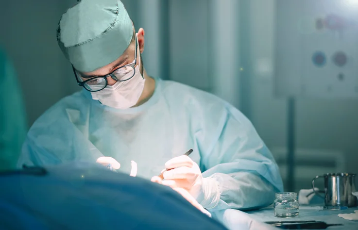
In the high-stakes environment of the operating room, where every millisecond counts and anatomical variations can turn a routine procedure into a complex challenge, information is the most valuable tool a surgeon possesses. While traditional surgical planning has long relied on two-dimensional scans and mental visualization, a seismic shift is underway. Today, surgeons are stepping away from the flat screen and into a three-dimensional world, utilizing hyper-accurate models, virtual reality (VR) simulations, and patient-specific reconstructions to navigate the human body with unprecedented precision.
As noted by Dr. Justin Ryan, director of the Helen and Will Webster Foundation 3D Innovations (3DI) Lab at Rady Children’s Health San Diego, this evolution is fundamentally changing the surgical landscape. By transforming complex medical imaging into tangible assets, the medical community is moving toward a future where "on-the-fly" surgical adjustments are replaced by meticulous, data-driven foresight.
The Chronology of a Digital Transformation
The integration of 3D technology into healthcare is not a sudden phenomenon but the culmination of nearly 30 years of incremental progress.
- The 1990s: The Dawn of Medical Modeling. Initial attempts to bridge the gap between diagnostic imaging and physical representation began in the mid-1990s. Early pioneers utilized rudimentary rapid-prototyping machines to create basic anatomical models, though the technology was largely relegated to research settings due to high costs and slow processing speeds.
- The 2000s: Refinement and Software Development. As software for CT and MRI segmentation became more sophisticated, the ability to translate "DICOM" data—the global standard for medical imaging—into printable formats improved. This decade saw the first successful clinical applications in maxillofacial and orthopedic surgery.
- The 2010s: The Proliferation of In-Hospital Labs. As printer costs plummeted and reliability surged, major academic medical centers began establishing dedicated 3D printing labs. This era marked the transition from "experimental research" to "standard of care" for complex congenital heart disease and neurosurgery.
- The 2020s: The Era of Immersive Media. Today, the focus has shifted beyond mere physical models. The current frontier involves Virtual Reality (VR) and Augmented Reality (AR), allowing surgeons to "walk through" a patient’s vascular system before making a single incision.
Building the Patient-Specific Blueprint
The magic behind this transformation lies in a process known as segmentation and reconstruction. It begins with the patient’s own diagnostic data—typically CT scans or MRIs.
From Pixels to Physicality
Medical imaging provides a massive volume of raw data. A 3D modeling specialist uses specialized software to "segment" these images, essentially separating the anatomy of interest (such as a tumor, a valve, or a fractured bone) from the surrounding tissue. This digital reconstruction can be finished in minutes, allowing for immediate visualization.
If a physical model is required, the digital file is sent to an additive manufacturing suite. High-end medical 3D printers can produce complex, multi-material models—some mimicking the density of bone, others the elasticity of heart tissue—within a 24-hour window. These models allow surgeons to hold the patient’s specific anatomy in their hands, rotating it, examining hidden angles, and even practicing the physical maneuvers required for the surgery.
The Role of Custom Guides
Beyond static models, specialists are now creating surgical templates and guides. These are custom-fitted tools designed to snap onto a patient’s anatomy, featuring precise "depth markers." These markers tell the surgeon exactly where to drill or cut, effectively eliminating the guesswork that often accompanies unconventional, complex anatomical structures. By pre-bending tools to match these guides before entering the OR, surgeons can shave critical time off procedures.
Supporting Physicians and Patients: The Structural Challenge
Despite the clear benefits, the implementation of 3D technology faces a scaling hurdle. Currently, two primary models exist for integrating these technologies into a hospital:
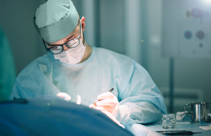
- The "Enthusiast" Model: In many facilities, 3D printing is championed by a sole, highly motivated surgeon or a small team. While this leads to exceptional outcomes for the patients of those specific doctors, it creates a "silo" effect. The technology does not permeate the rest of the hospital, leaving other departments—and their patients—without the benefits of these innovations.
- The "In-House Service Line" Model: This is the gold standard. A dedicated, in-house 3D lab acts as a hospital-wide resource. By centralizing the expertise, the hospital can foster a culture of collaboration. Specialists in the lab can act as consultants, working with neurosurgeons in the morning and cardiothoracic surgeons in the afternoon. This setup also allows for the "customization of the medium"—some surgeons may prefer a VR walkthrough, while others require a physical model to practice a suturing technique.
Bridging the Communication Gap
Perhaps the most overlooked benefit of 3D modeling is its role in patient and family communication. Surgery is inherently frightening, and explaining a complex procedure through a 2D scan is often ineffective. A color-coded, 3D-printed model of a child’s heart, for example, allows a surgeon to explain the pathology and the surgical plan in simple, visual terms. This empowers families, reduces anxiety, and builds trust, which is a vital component of the clinical process.
Supporting Data: Efficiency and Outcomes
The value proposition of 3D modeling is not merely qualitative; it is quantitative. Data from early adopters and hospital-wide initiatives suggest that the integration of 3D technologies can reduce operating room time by anywhere from 30 to 90 minutes per procedure.
- Cost Savings: While the initial setup of a 3D lab requires an investment in software, hardware, and specialized staff, the downstream savings are significant. Shorter surgeries mean reduced anesthesia time, lower risk of infection, faster recovery, and more efficient turnover of OR suites.
- Risk Mitigation: The "pre-op dress rehearsal" provided by VR or 3D models allows surgeons to identify potential complications—such as an artery that is positioned unexpectedly—before the first incision is made. This "measure twice, cut once" approach is fundamentally changing the risk profile of high-complexity surgeries.
Official Perspectives: The Path Forward
Dr. Justin Ryan, a leader in the field, emphasizes that the future of this technology will not be defined by the printers themselves, but by the processes surrounding them. "The technology is ready," he suggests. "The challenge now is organizational adoption."
The Radiological Society of North America (RSNA) has already begun standardizing best practices, ensuring that 3D printing in healthcare meets rigorous clinical appropriateness and quality assurance standards. As these standards become more universal, the barrier to entry for smaller hospitals will decrease.
However, the human element remains paramount. The success of 3D modeling in medicine depends on the collaboration between biomedical engineers and clinicians. It requires a shift in hospital culture—one that prioritizes interdisciplinary communication and embraces the "gaming" aspects of modern medicine, such as manipulating 3D reconstructions in virtual space as easily as one would a digital character.
Implications and The Future Horizon
As we look to the next decade, we can expect the integration of "4D" modeling—incorporating the dimension of time to show how an organ moves during a heartbeat or a breath. We will likely see the rise of "digital twins," where a patient’s entire physiological system is reconstructed to test the potential outcomes of various surgical interventions before the patient is even wheeled into the hospital.
The ultimate implication of this technological advancement is a more personalized medicine. We are moving away from a "one-size-fits-all" approach to anatomy and surgery. By embracing 3D modeling and immersive media, hospitals are not just upgrading their equipment; they are upgrading their capability to care for the individual.
In conclusion, the revolution of immersive media in the operating room is about more than just novelty. It is about equipping surgeons with the clarity they need to perform at their best, reducing the burden on patients, and creating a more efficient, safer healthcare system. While the technology is sophisticated, its goal is simple: to ensure that when a surgeon picks up a scalpel, they are acting with the highest possible level of knowledge, precision, and confidence. The hospitals that adapt to these new, integrated workflows will lead the next century of medical excellence, ultimately ensuring that we can support our physicians, so they can better support our patients.