








For the estimated millions of people living with Ehlers-Danlos syndrome (EDS), the journey to a diagnosis is often paved with frustration. A patient may present to their physician with debilitating neck pain, radiating neurological symptoms, or severe fatigue, only to undergo a standard MRI that returns the all-too-familiar result: "Unremarkable."
While this clinical finding provides relief to those without underlying pathology, for the EDS patient, it represents a diagnostic dead-end. The symptoms persist, yet the evidence remains elusive. As the medical community continues to refine its understanding of connective tissue disorders, a critical question has emerged: Are we looking at the right picture, or are we simply using the wrong frame? This article explores the growing interest in upright, weight-bearing MRI as a tool to bridge the gap between patient experience and objective clinical data.
The Diagnostic Mismatch: Why Supine MRI May Miss the Mark
To understand the utility of upright imaging, one must first understand the limitations of the "Gold Standard." The vast majority of diagnostic imaging performed globally uses high-field (1.5T or 3.0T) MRI scanners. These machines are designed for the patient to lie supine—flat on their back—in a static, gravity-neutralized position.
The Physics of the Problem
For most orthopedic and neurological conditions, supine imaging is perfectly adequate. However, EDS is a systemic disorder of connective tissue that frequently results in joint laxity and ligamentous instability. In these patients, the spine is not merely a static stack of bones; it is a dynamic structure held together by tissues that lack the structural integrity required to maintain alignment under the force of gravity.
When an EDS patient stands or sits, the weight of the head and torso exerts pressure on a spine that may already be compromised. If the ligaments are too loose to stabilize the vertebrae, the spine may shift, compress, or misalign—a phenomenon often called "dynamic instability." When that same patient is placed in a horizontal, supine position, gravity is removed from the equation. The vertebrae shift back into a neutral position, the spinal canal expands, and the very pathology causing the patient’s symptoms disappears from the view of the scanner.
This creates a "diagnostic mismatch." We are testing the spine in its most stable, least symptomatic state, while expecting to identify a mechanical problem that only manifests under the stress of daily living.
The Role of Positional (Upright) MRI
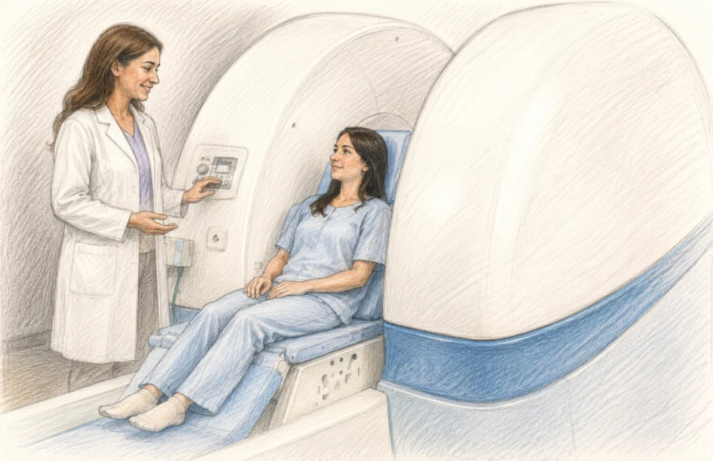
Positional MRI (pMRI), often referred to as weight-bearing or upright MRI, is a diagnostic modality designed to capture the anatomy under the physical demands of gravity. Unlike traditional machines, these scanners allow the patient to be imaged while sitting or standing. Some advanced systems even permit the patient to move into flexion, extension, or rotation, providing a "cinematic" view of how the spine behaves in motion.
The Trade-off: Resolution vs. Reality
Critics of upright MRI often point to a significant technological trade-off: field strength. Conventional hospital scanners typically operate at 1.5T to 3.0T, providing high-resolution, sharp images. Conversely, upright MRI systems generally operate at lower field strengths, typically ranging from 0.6T to 1.0T.
While this results in lower signal-to-noise ratios and potentially less crisp imagery, proponents argue that the utility of the image outweighs its aesthetic quality. If a 3.0T scan shows a "normal" spine while the patient is lying down, but a 0.6T upright scan reveals a 5mm shift in the vertebrae that compresses the spinal cord, the lower-resolution image is objectively more diagnostic for that specific patient.
Chronology of Clinical Interest: From Anecdote to Evidence
The interest in dynamic imaging for connective tissue disorders did not appear overnight. It is the result of decades of clinical observation.
- Early 2000s: Clinicians specializing in neurosurgery and genetics began noting a subset of EDS patients whose clinical symptoms—such as headaches, dizziness, and motor weakness—mirrored those of Chiari malformations and craniocervical instability (CCI).
- 2007: Landmark research by Milhorat et al. documented the syndrome of "cranial settling" in EDS patients, suggesting that the craniocervical junction was a primary site of mechanical failure. This brought weight-bearing imaging to the forefront of neurosurgical discussions.
- 2015: Health Quality Ontario released a systematic review evaluating the role of pMRI. While they acknowledged the difficulty in standardizing results, they highlighted the potential for revealing posture-dependent abnormalities that remain hidden in standard protocols.
- 2022–2025: A surge in systematic reviews, including the work of Lohkamp et al. and the 2025 scoping review by Verderame et al., established a more rigorous framework for evaluating spinal stability. These studies moved the conversation from "anecdotal" to "evidence-based," calling for standardized measurement criteria.
- 2026: The release of the "Manjila Chiari Protocol 2.0" (MaChiP 2.0) integrated AI-assisted analysis with dynamic imaging, signaling a shift toward precision medicine in the evaluation of complex head and neck symptoms in EDS.
Supporting Data: What Does the Research Say?
The primary strength of upright MRI lies in its ability to detect "occult" findings. Research consistently identifies several key abnormalities that are exacerbated by the upright position:
- Spinal Alignment Changes: Patients with EDS often show "subluxation" or sliding of vertebrae that only appears when the spine is loaded.
- Canal Narrowing: The diameter of the spinal canal can fluctuate significantly based on posture. An upright scan may reveal that the spinal cord is being pinched by ligaments or bone spurs that are not apparent while supine.
- CSF Flow Dynamics: In cases of Chiari malformation or suspected CSF leaks, upright imaging can show how the brain and spinal cord move relative to the skull, providing critical data on fluid dynamics that standard imaging misses.
- Tethered Cord Syndrome: Some studies indicate that postural changes can alter the tension on the spinal cord, particularly in patients with suspected occult tethered cord, a condition occasionally seen alongside hypermobility disorders.
Official Responses and Medical Guidelines
The medical community remains cautious but increasingly open to the role of pMRI. Organizations like the CCI Foundation have begun advocating for the inclusion of upright imaging as a supplementary tool in the diagnostic process for craniocervical instability.
However, major radiological bodies emphasize that upright MRI should not be viewed as a "silver bullet." The consensus among many neuroradiologists is that upright MRI should be reserved for cases where:
- Symptoms are strictly positional (e.g., headache improves when lying down, worsens when standing).
- Standard MRI is normal, but the clinical suspicion of instability remains high.
- The patient has a confirmed diagnosis of a connective tissue disorder and is experiencing new, progressive neurological deficits.
Implications for Patients and Clinicians
The adoption of upright MRI carries significant implications for the future of EDS care.
For the Patient
The primary implication is the validation of symptoms. For a patient who has spent years being told their "scans look fine," receiving an image that clearly shows the mechanical source of their pain can be life-changing. It provides a clear target for surgical or conservative interventions. However, patients must be prepared for the reality of "diagnostic ambiguity"—even an upright MRI may not provide a definitive answer, as the interpretation of "instability" is still being standardized across the medical community.
For the Healthcare System
Access remains the biggest hurdle. Upright MRI machines are expensive to maintain and require specialized training to interpret. Currently, these facilities are concentrated in major urban centers, forcing many patients to travel long distances or pay out-of-pocket, as insurance providers often deem these tests "investigational" or "not medically necessary."
As research grows, we expect to see:
- Standardized Protocols: Development of uniform criteria for what constitutes "pathological" movement in the spine.
- Insurance Parity: Increased coverage as more peer-reviewed studies validate the cost-effectiveness of getting the right diagnosis sooner.
- AI Integration: Using machine learning to measure the movement of the spine across thousands of frames, allowing for more precise calculations of instability than the human eye can perform alone.
When Might Upright MRI Be Relevant for You?
If you are navigating the complexities of EDS, you may want to discuss the following with your specialist:
- Positional Symptoms: Do your symptoms improve significantly when you lie down?
- Neurological Deficits: Are you experiencing new numbness, tingling, or weakness that correlates with specific head or neck movements?
- Discrepancies: Does your physical examination suggest instability (e.g., hyperreflexia or motor weakness) that is not reflected in your "unremarkable" supine MRI?
- Pre-surgical Planning: If surgery is being considered, does your surgeon have a complete understanding of how your spine behaves under gravity?
Key Takeaways
- The Spine is Dynamic: EDS affects the integrity of the structures holding the spine together, making it inherently sensitive to gravity and position.
- Diagnostic Mismatch: Standard MRI (supine) may miss instability that only occurs when the body is upright.
- A Tool, Not a Replacement: Upright MRI is a supplementary tool to be used when clinical symptoms contradict conventional imaging.
- The Need for Research: While promising, more large-scale studies are required to establish universal diagnostic criteria for EDS-related spinal instability.
- Advocacy and Access: Patients should work closely with their medical teams to determine if the benefits of positional imaging justify the cost and logistical challenges of accessing the technology.
In the final analysis, the goal of modern medicine is to match the diagnostic tool to the underlying physiology of the disease. For Ehlers-Danlos syndrome, that means acknowledging that the spine is not a static object—it is a structure in constant motion. By refining our ability to see that motion, we move one step closer to ensuring that no patient is left without answers.
Amy Weintraub is a Research Specialist II at the Norris Lab. This article was edited by Jacqueline Teti, Editor-in-Chief. Published June 2026.