
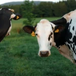


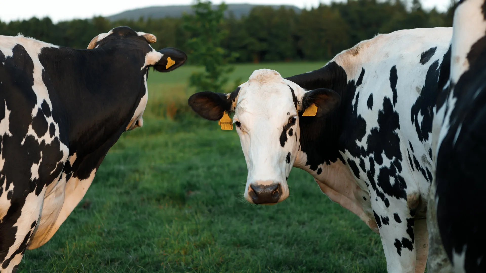
When the first reports of a mysterious illness began trickling out of the Texas Panhandle in early 2024, the dairy industry was braced for the usual culprits. Veterinarians, conditioned by decades of experience with bovine health, were on the lookout for common bacterial pathogens—the typical agents of mastitis, an inflammatory condition that causes severe distress in milk-producing animals. What they found instead was a viral anomaly that defied established veterinary science, signaling the arrival of a pathogen that did not play by the rules: the H5N1 strain of Highly Pathogenic Avian Influenza (HPAI).
Unlike previous iterations of influenza that wreak havoc on the respiratory systems of mammals, this virus took an unprecedented detour, anchoring itself firmly within the mammary glands of cattle. Now, a groundbreaking study from the University of Pittsburgh School of Public Health, published in Science Advances, has finally unlocked the biological mystery of why this virus chose the udder over the lungs, providing a critical roadmap for future pandemic preparedness.
The Chronology of an Unseen Invasion
The emergence of H5N1 in U.S. dairy cattle represented a significant paradigm shift in zoonotic disease. Historically, bird flu was considered a respiratory threat. However, the 2024 outbreak displayed a clinical profile that caught the global scientific community completely off guard.
The Initial Misidentification
In the early weeks of the outbreak, clinicians focused on standard bacterial mastitis. Because the affected cows did not exhibit the typical sneezing, coughing, or lethargy associated with respiratory flu, the idea that an influenza virus was the root cause was not even a remote consideration. This delayed identification allowed the virus to circulate silently through herds, turning milk—a primary product of the dairy industry—into a vehicle for viral shedding.
The Realization
As the virus continued to spread, the sheer volume of H5N1 present in the raw milk of infected cattle became clear. It was this discovery that shifted the narrative from a localized animal health issue to a broader public health concern. The realization that the virus was not just present, but thriving in the mammary tissue, forced a rapid pivot in surveillance strategies. Researchers began to track not only the movement of the cattle but the environmental contamination left in their wake, identifying a cycle of transmission that necessitated immediate intervention.
Unmasking the Culprit: The Glycan Architecture
At the heart of the University of Pittsburgh’s research is the question of "receptor biology." Influenza viruses are notoriously picky; they cannot simply invade any cell they encounter. Instead, they must perform a molecular "handshake" with specific receptors on the surface of host cells. These receptors, known as glycans, are sugar-based molecules that act as a lock, while the virus carries the key.
The Receptor Discrepancy
Previous studies had suggested that the glycan receptors typically favored by avian influenza were present in the noses, tracheas, and lungs of cattle. Theoretically, this should have made cows susceptible to a respiratory infection. Yet, the clinical reality showed the exact opposite: the lungs remained largely clear, while the udders were under siege.
To bridge this gap, Dr. Suresh Kuchipudi, chair of Infectious Diseases and Microbiology at Pitt Public Health, partnered with Dr. Lauren E. Pepi of Harvard Medical School, an expert in glycomics. The team realized that the standard models were too simplistic. "Glycan biology is very complex," Dr. Kuchipudi explained. "We realized that to understand what was really going on, we would need to use more innovative technologies and map out the fine-detailed architecture that enables the virus to bind to cells."
The "Perfect Breeding Ground"
By employing a sophisticated array of binding experiments, histochemical staining, and ultra-high-resolution imaging, the team identified a specific subtype of receptor: the N-linked sialic acid receptor. Their findings revealed that these specific receptors were abundantly distributed throughout bovine udder tissue but were nearly non-existent in the airway tissue.
This discovery provided the "smoking gun." The virus was not ignoring the cow; it was merely restricted to the specific biological niche where its required "lock" was available. The mammary glands, rich in these N-linked sialic acid receptors, became the "perfect breeding ground" for H5N1, explaining the severe necrotizing mastitis observed in the field.
Supporting Data and Clinical Implications
The implications of this research extend far beyond bovine health. The study serves as a masterclass in how viruses evolve to exploit the unique biological niches of different species.
The Occupational and Zombified Risks
The high viral load found in the milk of infected cows introduced a multi-tiered risk profile:
- Occupational Hazard: Farm workers in direct contact with infected udders and milk faced a heightened risk of exposure, a primary focus of federal and state health agencies.
- Domestic Transmission: The practice of feeding raw milk to domestic pets, such as cats, proved fatal in several documented instances. Research into these cases confirmed that the virus could cause severe neurological damage in felines, a stark contrast to the mastitis-driven symptoms in cattle.
- The Pasteurization Defense: Dr. Kuchipudi and his team were quick to reinforce a critical public health message: pasteurization is a highly effective, time-tested process that destroys the virus. The danger is not inherent in milk itself, but in the consumption of raw, untreated products.
Data-Driven Surveillance
The methodology developed at the University of Pittsburgh offers a new tool for global biosurveillance. By mapping the glycan receptor landscape of various species, scientists can now predict which animals might act as "intermediate hosts" for future influenza strains.
Official Responses and Future Preparedness
The U.S. Department of Agriculture (USDA) and various public health institutions have utilized the findings from this study to refine their containment strategies. The realization that H5N1 could bypass respiratory defenses and establish itself in reproductive/mammary tissues has necessitated a change in how diagnostic testing is deployed.
"We can preemptively screen different species and different tissues within them for susceptibility," Dr. Kuchipudi noted. This proactive approach—shifting from reactive testing to predictive mapping—is the cornerstone of modern pandemic prevention. By understanding the "why" behind the tissue-specific infection, officials can now create more accurate risk assessments for livestock and wildlife.
A New Era of Predictive Virology
The H5N1 outbreak in U.S. dairy cattle served as a humbling reminder of the fluidity of viral evolution. It demonstrated that even well-studied viruses can find novel ways to persist in environments that were previously considered "low risk" for respiratory transmission.
The collaborative effort between the University of Pittsburgh, Harvard Medical School, and other participating institutions—including Pennsylvania State University and North Dakota State University—represents the gold standard of cross-disciplinary research. By combining the physical sciences of glycomics with the practical, on-the-ground reality of veterinary medicine, these researchers have provided the world with more than just an explanation for a localized outbreak. They have provided a template for understanding the next potential pandemic.
As researchers look toward the future, the lessons learned from the "udder-centric" H5N1 outbreak will likely inform the development of vaccines, targeted biosecurity measures, and rapid-response protocols. The goal is no longer just to fight the current infection, but to anticipate the next "lock-and-key" mechanism that a virus might exploit. In the face of a changing biological landscape, this scientific vigilance is the most effective tool we have to prevent being caught by surprise again.
Study Contributors:
The research was led by Dr. Suresh Kuchipudi and included contributions from Surabhi Srinivas, Shubhada K. Chothe, Santhamani Ramasamy, Sougat Misra, Noel Chandan Nallipogu, and Lindsey LaBella (University of Pittsburgh); Yin-Ting Yeh (Pennsylvania State University); May Wang (Harvard University); and Heidi L. Pecoraro and Brett T. Webb (North Dakota State University).
This research was supported by the University of Pittsburgh School of Public Health and the U.S. Department of Agriculture’s National Institute of Food and Agriculture (Grant #FP00039373/AWD00010780).