








In a discovery that challenges decades of conventional wisdom, researchers at the University of Kentucky have identified an unexpected biological culprit behind the chronic sleep disturbances associated with Alzheimer’s disease. For years, the scientific community has focused primarily on amyloid plaques—the sticky protein deposits that accumulate between neurons—as the root cause of the cognitive decline and sleep fragmentation observed in patients.
However, a new study published in the journal Alzheimer’s & Dementia suggests that the problem is not necessarily the plaques themselves, but rather the brain’s aggressive, inflammatory response to them. By temporarily depleting the brain’s resident immune cells, researchers were able to restore more than two hours of sleep per day in mice models, achieving these results without removing a single plaque. This paradigm-shifting finding offers a potential roadmap for new therapies that could treat one of the most debilitating symptoms of Alzheimer’s without the need for complex, often invasive amyloid-clearance treatments.
The "Whole House" Response: A New Theory of Inflammation
To understand the significance of this discovery, one must first reconsider the role of microglia. These specialized immune cells are the brain’s first line of defense; under normal conditions, they are tasked with clearing debris, responding to injuries, and maintaining a homeostatic environment for neurons.
Dr. Shannon L. Macauley, an associate professor of physiology at the University of Kentucky College of Medicine and the study’s lead investigator, offers a compelling analogy to explain what happens when this system goes awry. "Imagine a small fire breaks out in a kitchen," she explains. "Instead of containing the flames to that room, the building’s entire sprinkler system activates, flooding the entire house and causing far more damage than the original fire."
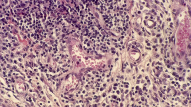
In the context of Alzheimer’s, microglia act like that overzealous sprinkler system. When they detect amyloid plaques, they don’t just clear the protein; they trigger a massive, sustained inflammatory cascade. This "whole house" defensive reaction creates a toxic environment that keeps the brain in a state of hyper-arousal, essentially preventing it from entering the deep, restorative stages of sleep.
"Microglia are immune cells that, when they respond to plaques, kick off this elaborate cascade of inflammation, as if the microglia are partying all night and keeping the brain awake," Dr. Macauley noted. The study’s first author, Dr. Nicholas J. Constantino, added that the discovery fundamentally alters how we perceive the progression of the disease, shifting the focus from the buildup of proteins to the brain’s reactive environment.
Chronology of Discovery: From Mice to Mechanism
The research team employed a multi-faceted approach to isolate the role of microglia in sleep pathology. They utilized two groups of mice: one group genetically predisposed to develop amyloid plaques and a control group of "wild-type" mice that aged naturally.
The study followed these subjects at two critical developmental milestones: six months, when the first plaques typically appear, and 18 months, when the Alzheimer’s-like pathology reaches an advanced stage. By comparing these groups, the researchers were able to isolate the specific sleep disruptions caused by Alzheimer’s from those naturally occurring as a byproduct of aging.
Tracking the Brain’s Electrical Rhythms
To monitor the animals, the team utilized advanced neuro-technologies. Each mouse was fitted with miniaturized devices capable of recording electroencephalography (EEG) and electromyography (EMG). While EEG tracked the brain’s electrical rhythms—revealing whether the mice were awake, dreaming, or in deep sleep—the EMG recorded muscle activity, providing a complete picture of the sleep-wake architecture.
To visualize the distribution of plaques and immune cells, the team utilized light-sheet microscopy. By making biological tissue transparent and illuminating it with laser light, the researchers created detailed, three-dimensional maps of the brain. This allowed them to see, with unprecedented clarity, exactly where the microglia were clustering in relation to the amyloid deposits.
The Intervention: Depleting the Immune Response
The pivot point of the research occurred when the team decided to test the effect of removing the microglia. Using a pharmacological agent called Pexidartinib (PLX3397)—a drug originally developed for oncology research—the team blocked the survival signals upon which microglia depend. After just 14 days of treatment, the researchers successfully depleted approximately 87% of the microglia in the mice brains.
The results were immediate and startling. The mice regained over two hours of sleep per day. Furthermore, the quality of that sleep improved, with the subjects spending significantly more time in Non-Rapid Eye Movement (NREM) sleep—the deepest, most physically restorative phase of the sleep cycle.
Supporting Data and the "Ceiling Effect"
One of the most counterintuitive findings of the study was the lack of correlation between the severity of plaque buildup and the intensity of sleep disruption. The researchers had hypothesized that as plaque burden increased, sleep would progressively worsen. Instead, they discovered a "ceiling effect."
"The disruptions in sleep and cortical EEG activity that occur at 6 months, when plaques first emerge, did not worsen by 18 months, despite more than double the amount of plaque burden," Dr. Constantino observed. This suggests that once the microglia reach a certain threshold of activation, they "max out" their disruptive influence on sleep. This discovery is crucial because it implies that sleep loss is an early-stage symptom of the disease, driven by the onset of inflammation rather than the sheer volume of protein accumulation.
Additionally, the study differentiated between the types of sleep loss caused by normal aging versus Alzheimer’s. While normal aging typically results in a decline in REM sleep (the stage associated with dreaming and memory consolidation), Alzheimer’s pathology was found to selectively sabotage NREM sleep. Given that NREM sleep is vital for clearing metabolic waste from the brain, this disruption creates a vicious, self-reinforcing cycle: the disease prevents the brain from cleaning its own toxins, and the buildup of those toxins further drives the disease.
Official Perspectives and Academic Collaboration
Dr. Macauley describes the project as a "beautiful partnership" built on a foundation of intellectual curiosity and risk-taking. Within the Department of Physiology at the University of Kentucky’s Sanders-Brown Center on Aging, the team fostered an environment where failing hypotheses were treated as necessary stepping stones rather than setbacks.
"I love people who take initiative, find their passion, are curious, and keep pushing to find an answer," Dr. Macauley stated. Her philosophy, summarized by the Wayne Gretzky quote, "You miss 100% of the shots you don’t take," served as the guiding principle for the laboratory.
The research was supported by a robust network of federal and private funding, including the National Institute on Aging (NIH), the National Institute of General Medical Sciences, the Cure Alzheimer’s Fund, and The CART Fund. This multi-institutional backing underscores the high level of confidence the scientific community has placed in this novel line of inquiry.
Implications: The Path Toward New Therapies
While the results in mice are groundbreaking, the researchers are careful to emphasize that they are not suggesting a clinical treatment that involves wiping out the immune system. Microglia are essential for healthy brain function, and their total removal is neither practical nor safe for human patients.
Repurposing Existing Drugs
The current focus of the team has shifted toward finding ways to "calm" overactive microglia rather than eliminating them. The lab is currently investigating existing medications—specifically the diabetes drug Metformin and the anti-seizure medication Stiripentol. These drugs are known to influence cellular metabolism, and researchers believe they may be able to tweak the metabolic energy of microglia, preventing them from entering that hyper-inflammatory "partying" state.
A New Diagnostic Tool: The Power of Portable EEG
Beyond treatment, the study highlights the potential for portable EEG devices to become a standard tool in Alzheimer’s screening. Because EEG records are non-invasive and significantly more affordable than MRI or PET scans, the team believes that these devices could eventually serve as "longitudinal biomarkers."
"Portable EEG systems could allow us to monitor people in their home environments and potentially screen for changes associated with an Alzheimer’s disease," Dr. Macauley suggested. For rural or underserved populations, this could be a game-changer, allowing for early detection without the need for expensive travel or invasive procedures.
Conclusion: A New Frontier in Alzheimer’s Research
The findings from the University of Kentucky represent a major shift in the Alzheimer’s landscape. By moving the spotlight away from the static plaques and onto the dynamic, inflammatory reaction of the brain’s immune system, the team has opened a new door for potential interventions.
If this model holds true for humans, the implications are profound: we may be able to improve the quality of life, cognition, and sleep for millions of patients by simply managing the "fire department" of the brain. While there is still a long road of clinical trials ahead, the message is clear: when it comes to the battle against Alzheimer’s, the solution may lie in the brain’s own response to the disease, rather than the disease itself.
This research, supported by various NIH awards and private foundations, continues to evolve, with the team now looking to translate their findings into human clinical studies.