


In the complex landscape of gastroenterology, few conditions pose as significant a diagnostic challenge as Chronic Ischemic Gastritis (CIG). Often described as a "clinical chameleon," CIG frequently mimics common gastric ailments, leading to delays in treatment that can escalate into life-threatening thromboembolic events. However, a recent case study involving a 63-year-old patient has provided a potential breakthrough, identifying a specific endoscopic visual marker—the appearance of evolving white patches on the gastric mucosa—that may serve as a critical red flag for clinicians.
This discovery is not merely a clinical curiosity; it represents a potential shift in how physicians approach patients with chronic, unexplained gastric ulcers and refractory epigastric pain. By linking these subtle endoscopic changes to underlying vascular compromise, the medical community may finally have a reliable, non-invasive "early warning system" to trigger definitive imaging before catastrophic complications occur.
Main Facts: The Diagnostic Conundrum of CIG
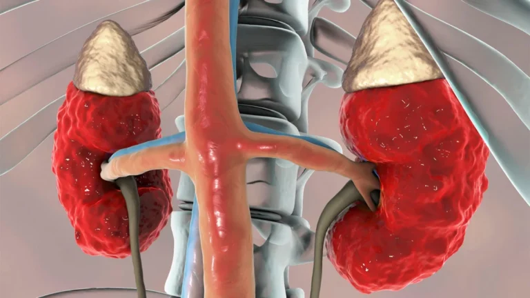
Chronic Ischemic Gastritis occurs when the blood supply to the stomach is chronically reduced, typically due to atherosclerotic disease of the major abdominal arteries, such as the celiac artery or the superior mesenteric artery (SMA). Because the stomach possesses a robust collateral blood supply, it is relatively resistant to acute ischemia. However, when multiple vessels are compromised, the stomach’s ability to maintain its mucosal barrier is severely undermined.
The Challenge of Recognition
Diagnosing CIG is notoriously difficult. Standard diagnostic tools—patient history, standard endoscopic examination, and even tissue biopsies—often yield ambiguous results. In many cases, CIG presents as common gastric ulcers or chronic gastritis, leading practitioners to treat the symptoms rather than the root cause: vascular insufficiency.
The core challenge lies in the fact that conventional biopsies often show non-specific inflammation or ischemic changes that are indistinguishable from common peptic ulcer disease. Consequently, clinicians are rarely prompted to order the necessary gold-standard diagnostic tool: abdominal CT angiography. This is where the identification of "white patches" becomes pivotal. This specific morphological change serves as a bridge, linking the visual findings of the endoscopist to the vascular assessment of the radiologist.
Chronology of a Case Study: From Pain to Recovery
The recent case that has gained attention in the medical literature provides a clear timeline of how this diagnostic marker can be utilized in practice.
Initial Presentation
A 63-year-old male presented to the clinic complaining of persistent, recurring epigastric pain. His medical history was marked by a series of unexplained gastric ulcers that had been treated multiple times without achieving long-term resolution. This "revolving door" of ulcer recurrence was the first subtle indicator that a systemic, rather than local, issue was at play.
The Endoscopic "Aha" Moment
During the upper endoscopy, physicians observed something distinct: the gastric mucosa was peppered with multiple, evolving white patches. Unlike standard ulceration or inflammatory plaques, these patches showed a progressive, temporal change. The endoscopist, suspecting that these patches were a visual manifestation of micro-vascular compromise—specifically, central vessel stenosis—ordered an immediate follow-up.
Vascular Investigation
Acting on the suspicion raised by the endoscopy, the medical team performed an aortic computed tomography (CT) scan. The imaging confirmed the clinical intuition: the patient exhibited complete occlusion of the superior mesenteric artery and significant stenosis of the celiac artery. The "white patches" were, in fact, localized areas of mucosal ischemia resulting from the severely restricted blood flow.
Surgical Intervention and Resolution
Following the diagnosis, the patient underwent a surgical bypass to restore blood flow to the affected region. The success of the procedure was confirmed not only by the patient’s immediate cessation of abdominal pain but, crucially, by follow-up endoscopy. The white patches had completely resolved, and the gastric mucosa had regained a healthy, uniform appearance, providing definitive proof that the mucosal changes were secondary to ischemia.
Supporting Data: Why "White Patches" Matter
The identification of these white patches as a diagnostic surrogate is supported by the underlying pathophysiology of CIG. The stomach’s mucosa is highly metabolically active and requires a consistent oxygen supply to maintain its structural integrity. When blood flow drops below a critical threshold, the superficial epithelial cells are the first to succumb.
The Pathophysiological Mechanism
The white patches are essentially "watershed" areas of the stomach. When blood flow is restricted, the tissue undergoes a process of coagulation necrosis or surface epithelial shedding, which manifests visually as a white, plaque-like appearance.
Supporting clinical literature suggests that:
- Temporal Evolution: These patches are not static; they change in appearance over time as the ischemia fluctuates, which explains why they might be missed in a single, isolated endoscopic view.
- Correlation with Vascular Anatomy: The patches often align with the distribution patterns of the celiac and mesenteric branches, providing a "map" of where the vascular insufficiency is most severe.
- Biopsy Limitations: While biopsies can show ischemic changes, they are often patchy and can be easily misidentified as simple H. pylori gastritis or autoimmune gastritis if the pathologist is not looking for vascular-specific indicators.
Official Responses and Clinical Perspectives
Medical experts and gastroenterologists have cautiously welcomed the report, noting that while the sample size is currently limited to case studies, the implications for clinical guidelines are significant.
"We have long struggled with the ‘cryptogenic’ ulcer patient," says one leading gastroenterologist. "When patients return repeatedly with ulcers that don’t respond to proton pump inhibitors or H. pylori eradication, we often blame patient compliance or aggressive acid production. This study forces us to look beyond the stomach and into the abdominal vasculature."
The Need for Interdisciplinary Collaboration
The consensus among experts is that this discovery underscores the necessity of a "vascular-first" mindset in patients with atypical gastric history. The official recommendation emerging from this case is that endoscopists should document not just the presence of ulcers, but the pattern and temporal changes of any mucosal irregularities. If white patches are observed in a patient with a history of cardiovascular disease, the threshold for ordering CT angiography should be significantly lowered.
Implications: Changing the Future of Gastric Care
The identification of white patches as a precursor to severe CIG could fundamentally change the management of gastric disease in the aging population.
Early Intervention as Life-Saving Care
CIG is not just a disease of the stomach; it is a manifestation of systemic atherosclerotic disease. The stomach serves as an early sentinel for the entire abdominal cavity. By diagnosing CIG through endoscopic observation, physicians can identify patients at high risk for mesenteric ischemia—a condition that, if left untreated, leads to bowel infarction, sepsis, and a high mortality rate.
Future Research Directions
The medical community is now looking toward larger, prospective studies to validate the "white patch" sign. Future research will likely focus on:
- Standardization: Creating an endoscopic scoring system for mucosal white patches to help clinicians distinguish them from other forms of gastritis.
- Artificial Intelligence Integration: Developing AI-assisted endoscopic software that can flag these specific patterns in real-time during a procedure, reducing the burden on the human eye.
- Longitudinal Tracking: Evaluating whether these patches correlate with the severity of stenosis in the celiac artery, potentially allowing for non-invasive "staging" of vascular disease via endoscopy.
A Paradigm Shift in Patient History
For the patient, this discovery means shorter diagnostic journeys. Instead of months or years of ineffective ulcer treatment, patients with these markers can be moved rapidly to vascular imaging and surgical consultation. It shifts the diagnostic focus from "Why is there acid?" to "Where is the blood flow?"
In conclusion, the case of the 63-year-old patient serves as a powerful reminder of the importance of clinical observation. By paying closer attention to the visual language of the gastric mucosa, gastroenterologists can catch a lethal disease in its early stages, preventing the progression from simple discomfort to life-threatening catastrophe. As the medical community integrates this "white patch" marker into standard practice, we may see a meaningful reduction in the morbidity associated with chronic ischemic gastritis, proving once again that the most sophisticated technology is often only as effective as the keen eyes of the clinician behind it.