










Long before the first cry of a newborn or the initial flickers of consciousness, the human brain undergoes an architectural transformation of staggering complexity. At the heart of this biological masterpiece lies a singular cell type: the radial glia. These specialized stem cells act as the master architects of the developing cerebral cortex—the seat of human thought, language, and memory.
For decades, the mechanisms by which these cells orchestrate the construction of our most complex organ remained largely shrouded in mystery. However, a pair of landmark studies published in the journals Cell and Science has fundamentally shifted our understanding. By integrating metabolic mapping and advanced neural "assembloids," researchers at the University of California, Los Angeles (UCLA) have revealed that radial glia are not merely biological blueprints; they are active, decision-making agents constantly recalibrating their work based on metabolic cues and physical touch.
The Foundation: Understanding Radial Glia
Radial glia are the progenitor cells from which the vast majority of our neurons arise. Their influence is so profound that they are widely believed to be the primary drivers of the evolutionary expansion of the human cortex. While other mammals possess similar cells, the human variant exhibits a unique capacity for persistence and proliferation, allowing for the creation of the massive, folded structure that distinguishes us from other species.
"Radial glia are the coolest cells that have ever existed," says Aparna Bhaduri, an assistant professor of biological chemistry at the David Geffen School of Medicine at UCLA. "They’re really key to making us human. But they’re also at the center of many neurodevelopmental and neuropsychiatric disorders, as well as cancer—so understanding how they make their decisions is one way to start understanding how those conditions arise."
The significance of these cells extends beyond prenatal development. In a cruel twist of biological irony, these same stem-like programs can be reactivated in brain cancers, leading to tumor growth. By deciphering the "decision-making" logic of radial glia, scientists are not just learning how the brain grows; they are unlocking a manual for potential therapeutic intervention in neurological disease and oncology.
Chronology of Discovery: A New Perspective on Development
For many years, the field of developmental biology operated under a relatively static model. Scientists assumed that the genetic code provided a rigid instruction manual and that the environment—metabolism and cellular interaction—played a secondary, "background" role.
Phase 1: The Metabolic Atlas (The Cell Study)
In the first phase of this research, led by co-first authors Jessenya Mil and Jose Soto, the team collaborated with the lab of Heather Christofk to create a high-resolution map of metabolic activity within the developing human cortex. Using donated tissue and brain organoids, the team made an unexpected discovery: metabolism is a command center, not a utility service.
The researchers identified that radial glia rely heavily on the pentose phosphate pathway. This metabolic route acts as a fuel-injection system, providing the necessary materials for rapid cell division. When the researchers experimentally restricted glucose or inhibited this pathway, the radial glia underwent a radical shift in output. Instead of producing the expected neurons, they began generating cell types that are usually reserved for later stages of brain development, effectively "rushing" or misaligning the developmental timeline.
Phase 2: The Physical Signal (The Science Study)
Simultaneously, a separate team led by Claudia Nguyen investigated the role of the thalamus. The thalamus, a deep-brain structure, sends long-distance projections to the cortex. Scientists have long observed these wire-like fibers arriving at the cortex surprisingly early—long before synaptic connections are finalized.
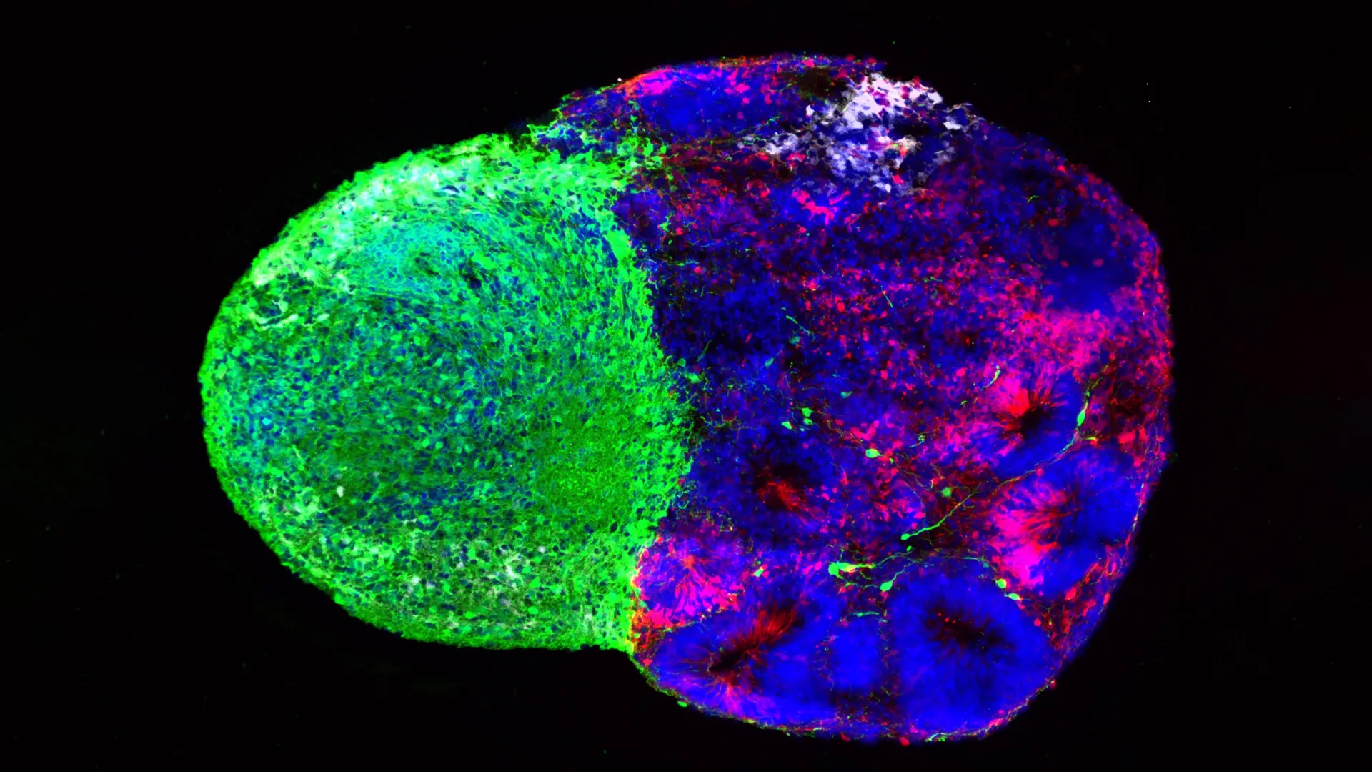
The researchers used "assembloids"—complex 3D models where different brain regions are grown together—to show that these thalamic fibers physically touch radial glia. This physical contact triggers a cascade of changes in the stem cells, prompting them to produce more excitatory neurons. These are the neurons responsible for carrying the primary signals through the cortex. This specific contact-based communication appears to be a uniquely human evolutionary development, absent in rodent models, which may explain the superior cognitive capacity of the human brain.
Supporting Data and Technical Breakthroughs
The success of these studies is inextricably linked to the rapid advancement of organoid technology. A decade ago, investigating the behavior of live human stem cells in a controlled environment was largely theoretical. Today, assembloids allow for the recreation of the "cross-talk" between different regions of the brain in a laboratory setting.
The data gathered from the metabolic atlas represents one of the most comprehensive resources ever compiled on brain development. By mapping the glucose consumption and metabolic flux of these cells, the team has provided a baseline that will allow future researchers to examine how external factors—such as maternal nutrition, metabolic disorders, or exposure to environmental toxins—can alter the "programming" of the fetal brain.
Furthermore, the team’s investigation into the gene NRXN1—a known risk factor for autism spectrum disorder—highlights the practical application of this research. By engineering assembloids with NRXN1 mutations, the team observed a breakdown in the physical signaling between the thalamus and the radial glia. This suggests that certain neurodevelopmental conditions may not be caused by a "broken" neuron, but by a failure in the early-stage communication between the brain’s structural components.
Official Responses and Expert Commentary
Dr. Aparna Bhaduri, who holds appointments at the UCLA Broad Stem Cell Research Center and the UCLA Health Jonsson Comprehensive Cancer Center, emphasizes that these findings represent a paradigm shift in how we view the "developing" brain.
"What was surprising is that metabolism isn’t just a passive thing that happens in the background," Bhaduri notes. "It can really control how stem cells make decisions. We already knew that thalamic projections influence how the cortex develops, but we specifically found that this influence comes through an actual physical connection—a point of contact that just hasn’t been identified before."
The research team suggests that these dual mechanisms—metabolic signaling and direct physical interaction—are likely working in concert throughout gestation. The brain is not just growing; it is constantly "listening" to its own internal environment, adjusting its trajectory based on the availability of nutrients and the connectivity of its constituent parts.
Implications for Future Research and Medicine
The implications of this research are far-reaching, touching upon neurology, psychiatry, and cancer research.
1. Neurodevelopmental Disorders
By understanding that radial glia decisions are modulated by signals like those from the thalamus, clinicians may eventually develop earlier screening tools for conditions such as autism or schizophrenia. If researchers can pinpoint the "timing" of these signals, they may eventually be able to identify developmental windows where the brain is most vulnerable to genetic or environmental disruptions.
2. Metabolic Health
The discovery that glucose levels influence cell fate suggests that the maternal metabolic environment is an even more active participant in fetal brain development than previously suspected. This underscores the importance of maternal health and nutrition, providing a clearer biological mechanism for how metabolic syndrome or gestational diabetes might affect long-term neurological outcomes.
3. Oncology
Perhaps most intriguing is the link to cancer. Since radial glia-like programs are often reactivated in malignant brain tumors, the metabolic pathways identified in these studies could become targets for new therapies. If a tumor is "mimicking" a developing brain to fuel its growth, then targeting the metabolic pathways that radial glia use could potentially starve the tumor without harming the healthy, mature brain tissue.
Conclusion: Looking Under the Hood
The human brain is a testament to the power of cellular coordination. By moving beyond the view of radial glia as static precursors, these two studies have opened a window into the dynamic, high-stakes environment of the fetal brain.
"Ultimately, these studies give us a glimpse under the hood of how these cells make decisions," Bhaduri concludes. "Understanding those decisions is a first step toward understanding normal brain development, disease vulnerability and, potentially, how similar stem-cell programs operate in brain cancer."
As organoid and assembloid technologies continue to mature, the ability to "see" into the earliest moments of human life will only grow sharper. For now, the work of the UCLA team provides the most detailed map to date of how we come to be, proving that our humanity is built not just on genes, but on a constant, complex dialogue between the cells that make us.
This research was supported by a wide coalition of scientific organizations, including the National Institutes of Health (NIH), the National Science Foundation (NSF), the Brain & Behavior Research Foundation, the Alfred P. Sloan Foundation, the Rose Hills Foundation, the Esther A. & Joseph Klingenstein Fund, the Simons Foundation, the Chan Zuckerberg Initiative, the NIH BRAIN Initiative Cell Atlas Network, the International Foundation for Ethical Research, and the UCLA Broad Stem Cell Research Center’s Ablon Scholars Program.