







In a landmark study that effectively rewrites the textbooks on human ocular development, researchers at Johns Hopkins University have decoded the precise biological choreography required to build the foveola—the infinitesimal center of the retina responsible for our sharpest, most detailed vision. By leveraging advanced organoid technology, the team has identified a sophisticated, two-step molecular switch involving vitamin A derivatives and thyroid hormones that transforms developing photoreceptors.
This discovery not only upends a 30-year-old scientific consensus regarding how the retina organizes its light-sensing cells but also provides a vital blueprint for future regenerative medicine. For the millions suffering from incurable degenerative conditions like macular degeneration and glaucoma, this research offers the first tangible glimmer of hope for "made-to-order" cell replacement therapies.
The Architecture of Sight: Why the Foveola Matters
To understand the magnitude of this discovery, one must appreciate the anatomy of human vision. The human retina is a complex, layered tissue at the back of the eye, populated by millions of light-sensitive neurons known as photoreceptors. These cells are divided into two categories: rods, which handle low-light and peripheral vision, and cones, which provide color and high-acuity daytime vision.
While the retina is vast, the foveola—a tiny depression at the very center of the macula—is the engine of our visual clarity. Despite occupying a minuscule fraction of the retinal surface, the foveola is responsible for roughly 50% of all visual processing in the human brain. Unlike the rest of the retina, which contains a mosaic of blue, green, and red-sensitive cones, the foveola is uniquely specialized: it is entirely devoid of blue-sensitive cones, housing only red and green receptors. This high-density, specialized arrangement is what allows humans to read, recognize faces, and perceive fine detail. Until now, the mechanism by which this unique "blue-free" zone was established remained one of ophthalmology’s most persistent enigmas.
Chronology of a Discovery: Tracking Fetal Development
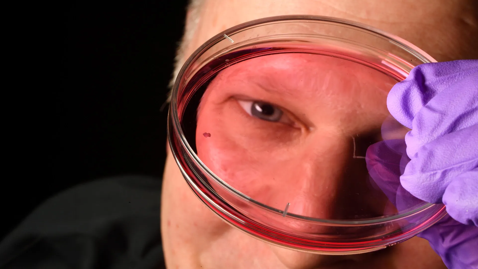
For decades, the study of human retinal development was hamstrung by a lack of suitable models. Common laboratory animals, such as mice and zebrafish, lack a foveola, making them poor proxies for human visual evolution. To bypass this, the Johns Hopkins team, led by Associate Professor of Biology Robert J. Johnston Jr., turned to retinal organoids—miniature, three-dimensional tissues grown from human fetal cells that mimic the physiological behavior of a developing eye.
The Developmental Timeline
By observing these organoids over several months, the researchers mapped the cellular events that define the foveola during critical windows of gestation:
- Weeks 10–12 (The Emergence): During the early stages of fetal development, the nascent foveola begins to produce blue-sensitive cones. At this stage, the region looks much like the rest of the retina, containing a mix of cone types.
- Week 14 (The Transformation): A dramatic shift occurs. The research indicates that the "blue-free" characteristic of the foveola is not achieved by the migration of cells, but by a process of conversion.
- The Molecular Triggers: The team identified that this conversion is governed by a two-part molecular signal. First, retinoic acid—a metabolite of vitamin A—is degraded, which halts the production of new blue cones. Second, thyroid hormones act upon the existing blue cones, physically re-engineering them into red and green cones.
"First, retinoic acid helps set the pattern," explains Johnston. "Then, thyroid hormone plays a role in converting the leftover cells. That’s very important because if you have those blue cones in there, you don’t see as well."
Challenging the Status Quo: Dismantling the Migration Theory
The findings published in the Proceedings of the National Academy of Sciences stand in direct opposition to a theory that has dominated the field since the early 1990s. The traditional model posited that the retina was essentially "hardwired" early on; it suggested that the foveola was initially populated by blue cones, which subsequently migrated away to the periphery, leaving a vacancy that was filled by red and green cones.
Johnston’s team has effectively debunked this "migration hypothesis." By tracking the cells in real-time within the organoids, the researchers observed that the blue cones do not vacate the center. Instead, they undergo a biological identity shift—a phenomenon known as cell plasticity—to become red and green cones. This discovery of "identity conversion" is a paradigm shift in developmental biology, suggesting that the retina is far more dynamic and malleable during fetal development than previously imagined.
Official Perspectives: The Path Forward
The implications of this research extend far beyond the laboratory. By uncovering the "instructions" the body uses to create the foveola, scientists can now attempt to replicate these conditions in a controlled environment to produce specific types of photoreceptors.
"This is a key step toward understanding the inner workings of the center of the retina, a critical part of the eye and the first to fail in people with macular degeneration," says Johnston. "By better understanding this region and developing organoids that mimic its function, we hope to one day grow and transplant these tissues to restore vision."
The research is particularly significant for Dr. Sarah Hussey, a former member of the Johnston lab and current molecular and cell biologist at CiRC Biosciences. Reflecting on the long-term clinical potential, Hussey emphasizes the methodical nature of translating organoid science into human therapy.
"The goal with using this organoid tech is to eventually make an almost made-to-order population of photoreceptors," Hussey notes. "A big avenue of potential is cell replacement therapy to introduce healthy cells that can reintegrate into the eye and potentially restore that lost vision. These are very long-term experiments, and of course, we’d need to do optimizations for safety and efficacy studies prior to moving into the clinic. But it’s a viable journey."
Future Implications: Restoring the Light
The vision of "made-to-order" photoreceptors could be the missing link in treating diseases like age-related macular degeneration (AMD). AMD currently affects millions of people globally, causing a gradual erosion of central vision that renders tasks like reading and driving impossible. Because the foveola is the specific site of degradation in AMD, the ability to regenerate these specific cells—or to provide a therapeutic substitute—would represent the "holy grail" of ophthalmology.
Hurdles and Next Steps
While the discovery is revolutionary, the team acknowledges that the path to clinical application is long. Future research will focus on:
- Refining Organoid Maturity: Ensuring that lab-grown tissue can reach a level of maturity that allows for successful integration into a living eye.
- Safety and Efficacy: Developing rigorous protocols to ensure that transplanted cells do not trigger immune rejection or develop uncontrolled growth.
- Integration: Solving the complex problem of "wiring"—ensuring that the new photoreceptors can effectively communicate with the existing neural network of the patient’s optic nerve and visual cortex.
Conclusion
The Johns Hopkins study is a masterclass in the power of modern biological modeling. By shifting the focus from observational, static studies of animal retinas to the active, developmental processes of human retinal organoids, Johnston and his colleagues have demystified one of the most complex structures in the human body.
While we are not yet at the stage of "growing eyes in a dish" for transplant, the identification of the retinoic acid and thyroid hormone switch provides the specific control mechanisms researchers have lacked for decades. As the field of regenerative medicine continues to accelerate, this work serves as a foundational pillar, bringing us one step closer to a future where blindness caused by retinal degeneration is no longer a permanent sentence, but a solvable biological problem.