







In the vast landscape of human anatomy, there is an implicit assumption that our internal organs are anchored securely in place, held by a sophisticated network of ligaments, fascia, and connective tissue. However, for a subset of the population—particularly those living with Hypermobile Ehlers-Danlos Syndrome (hEDS)—this structural integrity is often compromised. When these internal anchors fail, the result is visceroptosis, a condition defined by the downward displacement of abdominal organs.
While once a prominent topic of medical discourse at the turn of the 20th century, visceroptosis effectively vanished from the clinical lexicon for decades. Today, it is re-emerging, not as a curiosity of the past, but as a potential piece of the diagnostic puzzle for patients suffering from chronic, unexplained abdominal distress.
The Anatomy of Displacement: What is Visceroptosis?
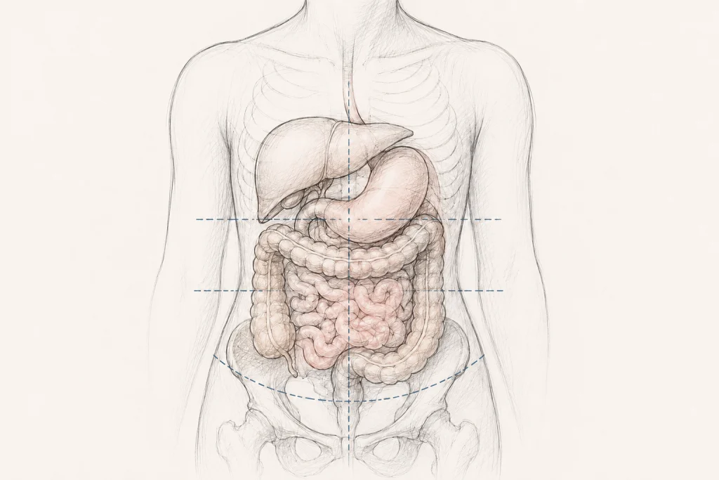
The term is derived from two Latin and Greek roots: viscera, referring to the internal organs of the abdominal and pelvic cavities, and ptosis, meaning a downward falling or displacement. In a healthy body, the stomach, liver, kidneys, and intestines are suspended by peritoneal attachments that maintain their position despite the forces of gravity.
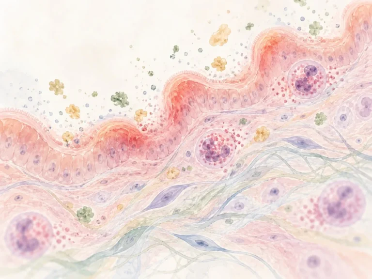
When those connective tissues are lax or weakened—a hallmark of systemic conditions like hEDS—these organs can descend significantly. This displacement is not merely a matter of anatomical aesthetics; it can fundamentally alter the physiological function of the organs involved. When the digestive tract, for instance, shifts downward, it may result in kinking, altered motility, or mechanical obstruction, leading to a host of debilitating symptoms that often defy conventional gastroenterological testing.
A Chronological Perspective: From "Glénard’s Disease" to Obscurity
To understand why visceroptosis is so misunderstood today, one must look back to the late 19th century.
The Era of Recognition (1885–1930s)
In 1885, French physician Dr. Frantz Glénard published landmark observations describing patients whose abdominal organs sat significantly lower than the accepted anatomical norm. At the time, "Glénard’s Disease" was widely accepted in medical journals like The New England Journal of Medicine, JAMA, and The Lancet. Radiologists and surgeons of the Victorian and Edwardian eras actively sought to identify and treat these displacements, viewing them as tangible physical pathologies.
The Great Disappearance
Sometime around the mid-20th century, visceroptosis suffered a quiet death in clinical practice. The medical community shifted its focus toward the emerging field of psychiatry and psychosomatic medicine. The "nervous" symptoms—anxiety, fatigue, and malaise—that often accompanied organ displacement were rebranded as purely psychological. Patients who complained of heavy, pulling sensations or chronic digestive failures were increasingly dismissed as "neurasthenic." The structural reality of their displaced organs was relegated to the fringes of medical textbooks.
The Modern Resurgence
Over the last two decades, the rise of awareness surrounding connective tissue disorders has forced a reckoning. Clinicians observing patients with hEDS have begun to note a high prevalence of organ prolapse. The clinical focus has moved from "nervous exhaustion" back to the connective tissue fragility that characterizes these genetic conditions, finally providing a biological framework for what early 20th-century physicians were seeing all along.
Supporting Data: The Link to Connective Tissue Laxity
The bridge between historical observations and modern science is being built through case studies and targeted research.
- Established Connections: Rectal and pelvic organ prolapse are already formally recognized within the 2017 diagnostic criteria for hEDS. This recognition validates that systemic laxity can, and does, lead to organ displacement.
- Gastrointestinal Evidence: A significant 2023 study involving 139 patients with slow-transit constipation identified a clear correlation between transverse colon ptosis and the severity of constipation. While this study did not focus exclusively on EDS, it provides robust evidence that anatomical descent is a major contributor to functional GI disease.
- The "Multifactorial" Hurdle: Current research faces a significant challenge: distinguishing between symptomatic visceroptosis and "anatomic" visceroptosis. As early as 1930, researchers warned that an organ sitting low is not, by itself, a disease. It only becomes "clinical visceroptosis" when that displacement directly interferes with function. In hEDS patients, this is complicated by concurrent issues like Autonomic Dysfunction (dysautonomia), disorders of gut-brain interaction, and pelvic floor dyssynergia, making it difficult to isolate the organ position as the sole cause of pain.
Clinical Implications: The Struggle for Diagnosis
The primary challenge for patients today is the lack of standardized diagnostic protocols. Standard gastrointestinal evaluations, such as endoscopies or colonoscopies, are designed to find lesions, inflammation, or tumors. They are not designed to measure the spatial position of organs in relation to gravity.
Because of this, many patients with visceroptosis are subjected to years of invasive testing that returns "normal" results, leading to a cycle of medical gaslighting. When a patient reports severe bloating, nausea, and early satiety—symptoms consistent with gastroparesis or severe constipation—the lack of a standard "visceroptosis scan" means the condition remains an invisible diagnosis.
Furthermore, there is a pervasive bias in the medical system where patients with chronic pain and systemic symptoms are often labeled as having "somatic" issues. This institutional skepticism remains the single greatest barrier to identifying and treating patients with clinically significant visceral displacement.
Treatment Paradigms: Navigating a Sparse Landscape
Treatment for visceroptosis remains in a transitional phase. Because there is no gold-standard surgical intervention for all forms of the condition, the approach is primarily conservative:
- Physical Therapy: Specialized pelvic floor and core-strengthening physical therapy is often the first line of defense. By stabilizing the musculature surrounding the abdominal cavity, patients may be able to support organs that lack proper ligamentous tethering.
- Compression and Support: The use of abdominal binders or compression garments is frequently reported by patients as a source of significant relief. These garments provide external structural support to the abdomen, which can help alleviate the "heavy" sensation associated with ptosis.
- Conservative Management: Dietary modifications to manage motility and the use of medications to aid digestion are standard.
- The Surgical Dilemma: Surgery for visceroptosis—such as colopexy or gastropexy—is a contentious topic. While successful outcomes are documented in literature, the surgical risks for hEDS patients are elevated due to tissue fragility and potential complications with healing. As such, surgery is generally considered a last resort, reserved for cases where mechanical obstruction is clearly identified and conservative measures have failed.
Moving Toward a New Standard of Care
The history of visceroptosis serves as a cautionary tale about how medicine can lose sight of structural pathologies by prioritizing psychological explanations. For the millions of people living with hEDS and other connective tissue disorders, the "forgetting" of visceroptosis has had real-world consequences, leading to decades of under-diagnosis and lack of targeted research.
As we move forward, the medical community must bridge the gap between early anatomical observations and modern imaging technology. Developing standardized, upright, or dynamic imaging protocols could allow clinicians to objectively measure organ displacement, moving the conversation away from anecdotal reports and toward evidence-based interventions.
Until that happens, the onus remains on patients and their providers to advocate for a more comprehensive view of abdominal pain—one that considers the structural integrity of the body’s internal architecture. The organs were meant to be held in place; when they aren’t, the consequences are anything but "all in the head."
Key Takeaways for Patients and Providers
- Validation: Visceroptosis is a documented medical phenomenon, not a psychological symptom.
- Context: It is frequently associated with systemic connective tissue disorders, including hEDS.
- Function vs. Anatomy: A diagnosis of clinical visceroptosis requires evidence that the displacement is causing functional impairment, not just that the organ is in a non-standard position.
- Advocacy: Patients should discuss the possibility of structural displacement with their specialists, particularly if their GI symptoms are accompanied by a sensation of "heaviness" or pressure that changes with position.
- Caution: Surgery should be approached with extreme caution in connective tissue patients due to the potential for poor healing and the complex nature of visceral symptoms.
Disclaimer: This article is for informational purposes only and does not constitute medical advice. Always seek the advice of a qualified healthcare provider with any questions regarding a medical condition.