











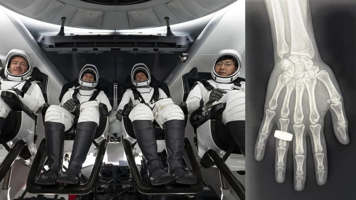
For over half a century, the history of human spaceflight has been defined by a narrow toolkit of medical diagnostics. When astronauts faced illness or injury in the vacuum of space, flight surgeons and mission commanders were largely limited to physical examinations and ultrasound imaging. However, a landmark experiment conducted during the Fram2 polar orbital mission in March 2025 has shattered this technological glass ceiling.
Researchers have successfully demonstrated that diagnostic-quality X-rays—a staple of terrestrial medicine for over a century—can be captured in the microgravity environment of Earth’s orbit. This achievement, detailed in the journal Radiology, represents a pivotal moment for the future of long-duration space travel and promises to yield significant secondary benefits for medical care in remote and austere environments back on Earth.
The Main Facts: Breaking the Microgravity Barrier
The core challenge of performing radiography in space has long been assumed to be insurmountable. Traditional X-ray systems are bulky, radiation-heavy, and notoriously sensitive to movement. In the microgravity of space, where subjects are constantly drifting and equipment lacks the stability of a solid floor, the potential for blurred or unusable images was long considered a disqualifying factor.
Led by Dr. Sheyna Gifford of the Mayo Clinic, a research team utilized a commercial-off-the-shelf (COTS) portable radiography system to conduct the first-ever human radiographs in orbit. The study involved three crew members on the Fram2 mission, a 3.5-day orbital flight launched via a SpaceX Falcon 9 rocket.
The results were unequivocal: non-medical professionals, after only four hours of training, were able to acquire high-quality diagnostic images of the human anatomy, including the chest, abdomen, pelvis, and extremities. While some minor positioning discrepancies were noted due to the lack of anchoring equipment, the diagnostic utility of the images was comparable to those taken in a controlled terrestrial environment.
Chronology: From Parabolic Simulation to Orbital Reality
The path to the Fram2 milestone was a calculated, multi-stage process that moved from theoretical design to extreme-environment testing.
Phase 1: The Parabolic Precursor
Before risking a launch, the team needed to understand how gravity fluctuations would impact X-ray output and image clarity. Dr. Gifford and her colleagues first conducted feasibility studies during parabolic flights—often referred to as the "Vomit Comet"—which simulate microgravity, lunar gravity, and Martian gravity. By capturing radiographs of both human subjects and specialized phantoms (test objects), the team proved that the physics of radiography could indeed function outside the standard gravitational pull of Earth.
Phase 2: The Training Protocol
Recognizing that space missions prioritize operational efficiency, the team designed a training program that was intentionally brief. Three crew members were selected for the project and underwent a total of four hours of instruction. This training covered the basics of radiation safety, system operation, and positioning. The goal was to prove that the system was intuitive enough for non-specialists to operate under the high-pressure, low-resource constraints of a space mission.
Phase 3: The Fram2 Mission (March 2025)
The critical test occurred during the Fram2 polar orbital flight. Upon reaching orbit, the crew deployed the portable system without any real-time guidance or ground-based support. They successfully imaged a variety of subjects, including a calibration phantom, a smartwatch, and various parts of their own bodies. These images were stored, reviewed, and transmitted to onboard computers, marking the first time humans had successfully performed their own diagnostic imaging while in orbit.
Supporting Data: Assessing Image Quality
To ensure scientific rigor, the team employed a blind evaluation process. Independent radiologists—specialists in abdominal and musculoskeletal imaging with years of clinical experience—reviewed the images. They utilized a Likert scale (1–5) to score the images based on clinical utility and clarity, comparing the in-flight results against preflight images taken on Earth.
Statistical Findings:
- Imaging Consistency: For most anatomical regions, the radiologists found no statistically significant difference in quality between Earth-based images and those captured in orbit.
- Positioning Challenges: The mean score for central radiographs (chest, abdomen, and pelvis) was 4.07 in-flight, compared to 4.95 preflight (P=0.02).
- User Feedback: All three crew members reported that the system was highly intuitive. However, the survey data highlighted a specific operational gap: the lack of anchoring hardware. In microgravity, keeping the X-ray detector and generator stable relative to the patient is difficult, and the crew noted that future iterations would require standardized clamping or mounting mechanisms to optimize patient positioning.
Despite the slightly lower score for positioning, the diagnostic quality remained high enough for clinical decision-making, confirming that the "conceit" that X-rays were too difficult for spaceflight had been effectively debunked.
Official Responses and Expert Perspective
The success of the experiment has drawn praise from the aerospace medicine community. Dr. Sheyna Gifford emphasized the simplicity of the achievement in a recent press release: "Acquiring diagnostically useful X-rays in space is something that anyone can do. Three very talented non-medical people with 4 hours of training in one of the harshest environments did it right and did it well."
The implications were further dissected in an editorial published alongside the study in Radiology by Dr. Suhny Abbara and Dr. Alan B. McMillan. They noted that the success of the project is not just a triumph for NASA or private spaceflight, but a fundamental shift in how we approach healthcare in remote locations.
"As our species crosses another boundary, from Earth to orbit and beyond, radiology is not merely following humanity into space; it is becoming part of the infrastructure that may make long-term exploration safer and more sustainable," the authors stated.
Implications: The Terrestrial Spillover
While the immediate goal of Dr. Gifford’s research was to keep astronauts safe during missions to the Moon or Mars, the long-term potential for Earth-based medicine is equally transformative. The "ruggedization" of medical equipment required for spaceflight is a well-trodden path for technological innovation.
1. Disaster Response and Military Medicine
In the wake of natural disasters or on the front lines of conflict, medical infrastructure is often the first thing to collapse. Portable, autonomous, and ruggedized X-ray systems could provide field medics with the ability to diagnose fractures, internal bleeding, or shrapnel injuries within minutes of arrival, without the need for a stable power grid or a hospital facility.
2. Remote and Austere Communities
Many rural communities around the world lack access to basic diagnostic imaging. A system that can be operated by a non-radiologist with minimal training could revolutionize care in underserved areas, turning local clinics into hubs for primary diagnostics that currently require hours of travel to a major city.
3. The Future of Exploration
As humanity looks toward long-term lunar bases and eventual crewed missions to Mars, the health of the crew becomes the most critical asset. A physician may not always be present on these missions; therefore, the ability for a crew to diagnose themselves using autonomous, guided imaging systems is not just a luxury—it is a mission-critical requirement.
Conclusion: Looking Ahead
The success of the Fram2 X-ray experiment serves as a reminder that the barriers to space exploration are often as much about our assumptions as they are about the physics of the cosmos. By proving that high-quality, actionable medical imaging is possible in microgravity, the team has taken a definitive step toward making space travel a sustainable endeavor.
Dr. Gifford and her team are now focused on the next stage of development: miniaturization and integration. As the technology continues to shrink in size and grow in reliability, the prospect of an "in-flight clinic" for long-duration missions moves from the realm of science fiction to reality. The lessons learned in the cramped, weightless environment of the Fram2 capsule will undoubtedly provide the foundation for the next generation of medical diagnostic tools, both in the stars and here on the ground.