










Influenza A remains one of humanity’s most persistent and formidable biological adversaries. Responsible for 3 to 5 million cases of severe illness and up to 650,000 deaths annually, its history is punctuated by devastating pandemics, most notably the 1918 “Spanish Flu.” Despite decades of research, the precise mechanics of how this virus converts a healthy human cell into a viral factory have remained partially obscured.
Now, a collaborative team from the European Molecular Biology Laboratory (EMBL) Hamburg and the Leibniz Research Institute for Molecular Pharmacology (FMP) has unveiled an unprecedented, high-resolution map of these interactions. By observing the virus inside intact, living cells—rather than the traditional, disruptive method of breaking cells apart—researchers have revealed the clandestine strategies influenza A employs to subvert human biological machinery. Published in Nature Microbiology, this study provides a blueprint for future antiviral therapies and a new paradigm for understanding viral pathogenesis.
The Chronology of an Infection
To understand the significance of this work, one must first understand the influenza life cycle. Upon entering a human cell, the virus releases its RNA payload, which contains the genetic blueprints for a small but powerful set of viral proteins. These proteins act as molecular saboteurs, infiltrating the cell’s internal communication and production networks to redirect resources toward the manufacturing of new virus particles.
For decades, the standard scientific approach to mapping these interactions involved “breaking open” the cell—a process known as cell lysis. While useful, this technique has a fundamental flaw: it destroys the cellular architecture. Proteins that were once physically separated by membrane walls or intracellular compartments may collide in the test tube, creating “false positive” interactions. Simultaneously, delicate, transient, or location-specific interactions—the very ones the virus relies on—often vanish during the chaos of cell destruction.
The current breakthrough began when the EMBL team, led by Jan Kosinski, joined forces with Boris Bogdanow and Fan Liu at the FMP in Berlin. They realized that a specialized technique, cross-linking mass spectrometry (XL-MS), could be optimized for the study of infected cells. By "freezing" protein interactions in place while the cell was still intact, the researchers were able to capture the virus in the act of hijacking, preserving the spatial context that is vital for understanding biological function.
Supporting Data: Integrating XL-MS and AlphaFold
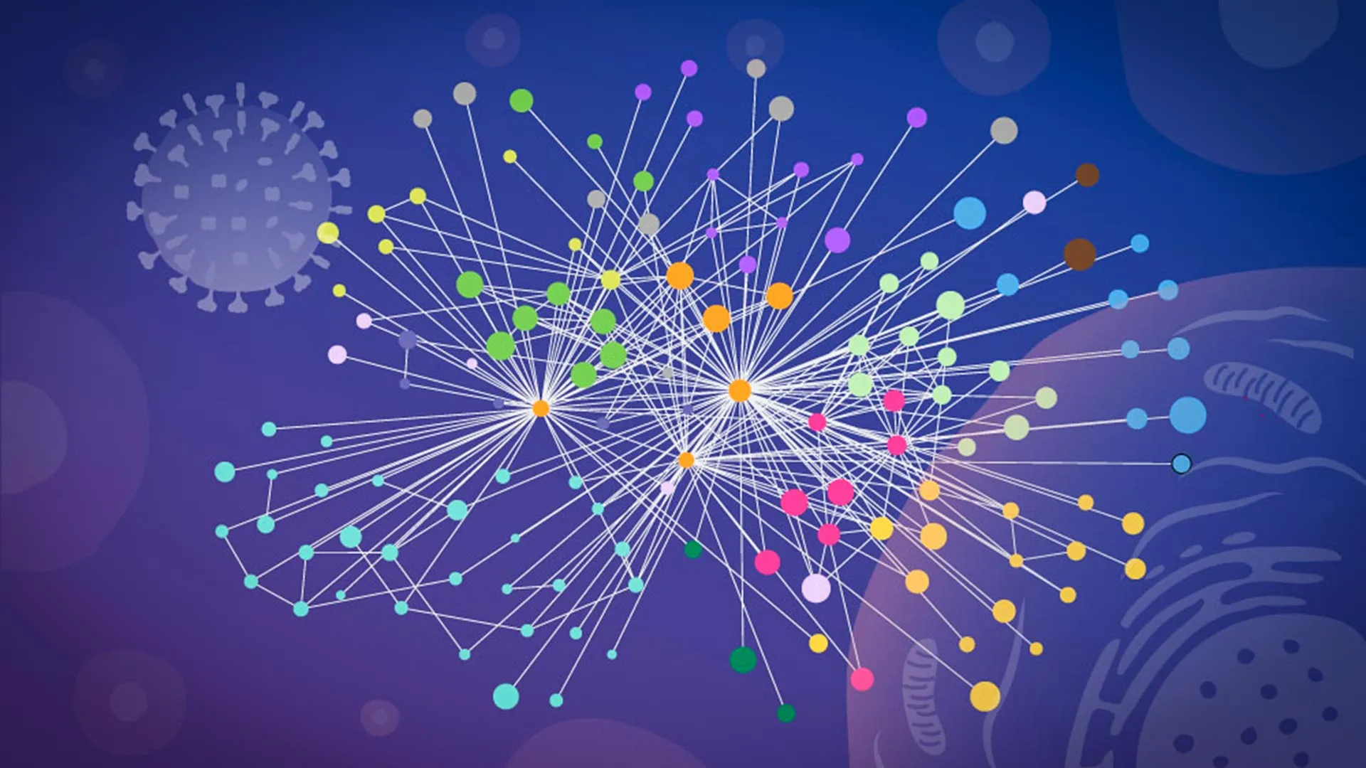
The success of this study hinged on the fusion of experimental data with cutting-edge computational modeling. The team utilized XL-MS to map the proximity of viral and host proteins, generating a vast dataset of physical contacts. To turn this raw data into a visual map, they turned to a modified version of AlphaFold—the Nobel Prize-winning AI algorithm capable of predicting protein structures.
“The key advantage of the modified AlphaFold approach is that it allowed us to feed our experimental cross-linking data directly into the structural modeling,” explained Jan Kosinski, Group Leader at EMBL Hamburg and the Centre for Structural Systems Biology (CSSB). By using the experimental data as a scaffold, the researchers could pinpoint which parts of the viral proteins were touching the host’s proteins, enabling them to build a 3D model of these interactions with unprecedented accuracy.
This structural detail is transformative. In pharmacology, knowing that a virus interacts with a human protein is only half the battle; knowing the exact "interface" where those two proteins lock together allows for the design of small-molecule drugs that can physically block that interaction, effectively stopping the virus in its tracks.
Two Strategies of Cellular Subversion
The study identified two distinct, high-impact strategies influenza A utilizes to secure its success within the host.
1. The Hemagglutinin Processing Network
The first strategy centers on hemagglutinin, the spike-like protein on the virus’s surface that allows it to latch onto and enter human cells. Once inside, hemagglutinin must be properly folded and modified before it can become part of a new virus particle. The researchers discovered that the virus hijacks the cell’s internal protein-processing network—the endoplasmic reticulum and the Golgi apparatus—to do this.
By mapping these interactions, the team identified specific human proteins that the virus forces to assist in the folding of hemagglutinin. Some of these host proteins were previously poorly understood, marking them as potential new targets for drugs that could prevent the virus from maturing.
2. The Dissolution of Paraspeckles
Perhaps the most surprising discovery involved "paraspeckles," small, droplet-like structures found inside the cell nucleus. These structures normally act as regulators for RNA and play a role in the cell’s stress response. The researchers observed that in every cell line and every flu strain tested, influenza A caused these paraspeckles to dissolve.
"Watching these tiny organelles in the nucleus dissolve, consistently across every cell line and every flu strain we tested, told us this isn’t a side effect of infection—it might be a strategy," said Iuliia Kotova, the study’s lead author and a former predoctoral fellow at EMBL Hamburg.
The dissolution of these organelles serves a dual purpose. First, it releases RNA-binding proteins that the virus then scavenges to support its own replication. Second, it cripples the cell’s ability to mount an effective antiviral defense, as paraspeckles are vital for regulating the cell’s stress response and immune signaling. By destroying these structures, the virus essentially "mutes" the cell’s alarm system.
Official Perspectives and Implications
The project was a tour-de-force of international collaboration, requiring the expertise of three major institutions. While the mass spectrometry took place at Charité – Universitätsmedizin Berlin, glycoproteomics was handled by the EMBL Proteomics Core Facility, and the complex structural modeling was processed on the EMBL Compute Cluster.
The implications for medicine are profound. By observing the virus in its native state, the researchers have provided a roadmap that moves beyond static snapshots toward a dynamic understanding of infection.
“Our work provides a new way to study flu-host interactions in their native context and with structural insight,” Kosinski stated. “The current results are a snapshot of a moment during infection, and it opens the door to studying flu-host interactions across the entire infection cycle.”
Boris Bogdanow, now a Junior Research Group Leader at the Institute of Virology at Charité, emphasizes the pharmaceutical potential: “This gives us insight into the interface between the virus and the human cell and may, through structural modeling, help identify actionable targets for future pharmaceutical interventions.”
Future Horizons: Beyond Influenza
While this study focused on a laboratory-adapted strain of influenza A, the methodology is designed to be universal. The researchers believe their “mapping in context” approach is a blueprint for studying any pathogen, including viruses with significant pandemic potential such as H5N1.
As global health authorities continue to monitor the threat of zoonotic viruses—those that jump from animals to humans—the ability to rapidly map how these pathogens interact with human biology is critical. By identifying the core proteins a virus must have to survive, scientists can develop “host-directed” therapies. Unlike traditional vaccines, which target the virus and can be rendered ineffective by viral mutations, host-directed drugs target the human proteins that the virus needs to replicate. Because these human proteins are less prone to mutation, such drugs could theoretically remain effective against a wide variety of viral strains.
In summary, the work of the EMBL-FMP team marks a departure from the reductionist biology of the past. By looking at the cell not as a collection of parts, but as a complex, living landscape, they have turned the spotlight on the virus’s most vulnerable moments. The map of the flu’s takeover is no longer a mystery; it is now a guide for the next generation of life-saving medicine.