











In the landscape of modern medicine, stress is often treated as an abstract, subjective phenomenon. While patients can report feelings of anxiety or fatigue, physicians have long lacked a concrete, objective way to measure the "cumulative toll" that chronic life stress takes on the human body. That is, until now.
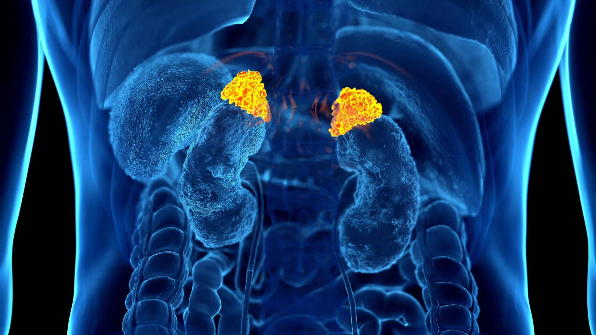
Researchers at the Johns Hopkins University School of Medicine have unveiled a breakthrough that promises to transform how clinicians view the physiological impact of chronic stress. By leveraging a sophisticated deep learning artificial intelligence model, the team has identified the first-ever imaging-based biomarker for chronic stress: the volume of the adrenal glands. This discovery, set to be presented at the upcoming annual meeting of the Radiological Society of North America (RSNA), turns the standard, widely performed CT scan into a powerful diagnostic tool for quantifying the biological "wear and tear" of a high-stress lifestyle.
The Physiology of the "Hidden" Stress Load
Chronic stress is far more than a psychological state; it is a systemic physiological burden. According to the American Psychological Association, prolonged exposure to stress hormones triggers a cascade of biological changes that can exacerbate anxiety, disrupt sleep patterns, induce muscle pain, drive up blood pressure, and suppress the immune system. Over years, this "allostatic load"—the cumulative physiological wear and tear on the body—is directly linked to the development of life-threatening conditions, including obesity, clinical depression, and cardiovascular disease.
Historically, measuring this load has been a logistical nightmare. While doctors can rely on patient-reported questionnaires, these are inherently subjective. Biological markers like cortisol levels in the blood or saliva provide a snapshot of stress at a specific moment in time, but they fail to capture the long-term, cumulative damage. Measuring cortisol is also notoriously cumbersome, often requiring multiple daily samples that are difficult to standardize.
The Johns Hopkins team, led by postdoctoral research fellow Elena Ghotbi, M.D., recognized that the answer might already be hidden in the terabytes of imaging data stored in hospital archives across the globe.
Chronology of a Medical Breakthrough
The development of this AI-driven biomarker was the result of a rigorous, multi-year validation process:
- Cohort Identification: The researchers utilized data from the Multi-Ethnic Study of Atherosclerosis (MESA), a massive, longitudinal study that provides a rare trifecta of data: high-resolution chest CT imaging, validated psychosocial stress questionnaires, and comprehensive biochemical markers of allostatic load.
- Tool Development: Dr. Ghotbi and her team engineered a deep learning algorithm specifically trained to identify, isolate, and calculate the volume of the adrenal glands on routine chest CT scans—a procedure performed tens of millions of times annually in the U.S. alone.
- Data Integration: The team established the "Adrenal Volume Index" (AVI), a metric that normalizes adrenal size by dividing volume by the patient’s height squared (cm³/m²). This was then cross-referenced against salivary cortisol levels (collected eight times a day over 48 hours) and indicators of allostatic load, such as body mass index, glucose levels, and blood pressure.
- Correlation and Validation: By comparing AVI scores against the 10-year health outcomes of the 2,842 participants in the MESA cohort, the team was able to draw direct, statistically significant links between enlarged adrenal glands and future adverse cardiovascular events.
Supporting Data: When Anatomy Meets Psychology
The data derived from this research provides the strongest evidence yet that our internal organs "keep the score" of our psychological state. The AI-driven analysis revealed that individuals with higher AVI values consistently showed greater overall cortisol exposure and higher peak cortisol levels.
More importantly, the research found a direct correlation between the AI-derived biomarker and physical heart health. Specifically, a higher AVI was linked to a higher left ventricular mass index—a structural change in the heart that is a precursor to heart failure. The study found that for every 1 cm³/m² increase in the Adrenal Volume Index, the risk of heart failure and mortality significantly increased.
This is the first time an imaging marker has been validated as an independent predictor of a major clinical outcome like heart failure. Unlike a single blood test, the size of the adrenal gland acts as a long-term "gauge" of chronic stress, reflecting a history of hormonal output that has physically reshaped the gland over time.
Official Responses and Expert Perspective
The implications of this study are being met with significant interest from the radiology and cardiology communities. Dr. Shadpour Demehri, a professor of radiology at Johns Hopkins and the senior author of the study, believes this discovery democratizes the ability to assess stress-related health risks.
"For the first time, we can ‘see’ the long-term burden of stress inside the body, using a scan that patients already get every day in hospitals across the country," Dr. Demehri stated. "Until now, we haven’t had a way to measure and quantify the cumulative effects of chronic stress, other than questionnaires or cumbersome serum markers. By connecting a simple imaging measure with well-established markers of stress, we are creating a new, practical approach for everyday clinical practice."
Dr. Teresa E. Seeman, a professor of epidemiology at UCLA and a pioneering researcher in the field of stress and health, emphasized the paradigm shift this work represents. "For over three decades, we’ve known that chronic stress can wear down the body across multiple systems," Dr. Seeman noted. "What makes this work so exciting is that it links a routinely obtained imaging feature with validated biological and psychological measures. It’s a true step forward in operationalizing the cumulative impact of stress on health."
Implications for Future Preventive Care
The most immediate implication of this study is the potential for improved cardiovascular risk stratification. Because the AI model can be applied to existing CT scans without the need for additional radiation or invasive testing, it could theoretically be integrated into routine radiological workflows immediately.
Enhancing Preventive Medicine
By identifying patients with elevated adrenal volumes, doctors could flag individuals at higher risk for cardiovascular events long before they manifest symptoms. This opens the door to early, targeted interventions—such as lifestyle changes, stress-reduction programs, or prophylactic cardiac care—that could prevent the onset of heart failure.
A New Diagnostic Standard
The Adrenal Volume Index could eventually become a standard measurement in radiology reports, much like a bone density score or a calcium score. This would allow physicians to track a patient’s "stress load" over time, providing a data-driven metric to see if lifestyle changes are effectively reducing the body’s physiological response to stress.
Applications Beyond Cardiology
While the current study focused heavily on heart-related outcomes, the researchers believe the utility of this biomarker extends further. Chronic stress is implicated in a wide range of diseases common in aging populations, from immune dysfunction to metabolic disorders. As the AI model is refined, it may eventually serve as a universal "stress score" that helps manage a variety of age-related conditions.
Conclusion: A New Lens on Health
The integration of artificial intelligence into medical imaging is no longer just about detecting tumors or fractures; it is beginning to map the very biological footprint of our modern lives. By turning the adrenal gland into a window through which we can observe the cumulative, invisible burden of stress, researchers have provided a vital new tool for preventive medicine.
As Dr. Ghotbi summarized, "Our approach leverages widely available imaging data and opens the door to large-scale evaluations of the biological impact of chronic stress. It has the potential to enhance risk stratification and guide preventive care without the need for any additional testing."
As the medical community gathers for the RSNA annual meeting, this study stands as a testament to how the marriage of deep learning and standard radiology can unlock deeper insights into the human condition, shifting the focus of medicine from merely treating the consequences of chronic stress to identifying and mitigating the damage before it becomes irreversible.