







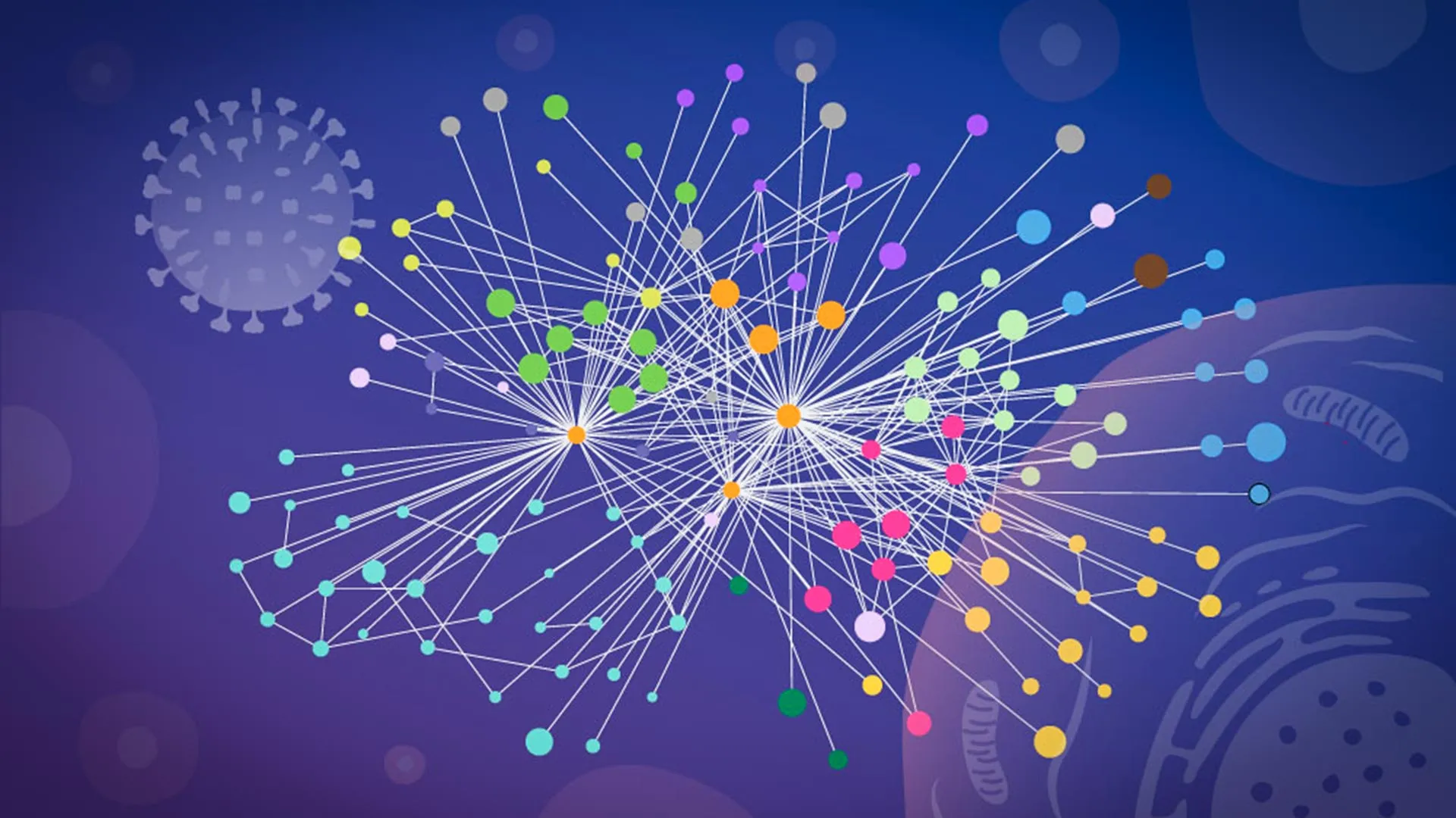
Influenza A, a viral adversary that causes 3 to 5 million cases of severe illness annually and claims up to 650,000 lives, remains a persistent global health threat. Beyond seasonal surges, the virus’s history of spawning devastating pandemics—most notably the 1918 Spanish Flu—has driven scientists to seek a deeper understanding of its survival tactics. A breakthrough study published in Nature Microbiology by researchers at EMBL Hamburg, in collaboration with the Leibniz Research Institute for Molecular Pharmacology (FMP) and Charité – Universitätsmedizin Berlin, has provided the most detailed map to date of how this virus systematically reshapes the human cellular environment.
By employing a revolutionary workflow that captures protein interactions within intact, living cells, the team has moved beyond the "broken-cell" limitations of the past, offering a high-resolution window into the molecular mechanisms of viral infection.
The Chronology of Cellular Takeover
To understand the significance of this new research, one must first understand the life cycle of the influenza virus. Upon entry into a host cell, the virus releases its RNA, which serves as a biological blueprint for viral proteins. These proteins act as a specialized invasion force, spreading throughout the host cell to redirect its internal molecular systems. Effectively, the virus transforms a healthy, functioning human cell into a biological factory dedicated to the mass production of new virus particles.
For decades, the challenge for virologists has been tracking this "takeover" in real-time. Traditional biochemical techniques required researchers to pulverize cells to measure protein contacts. However, this process is inherently destructive; once the delicate internal architecture of a cell is dismantled, proteins that were once sequestered in separate compartments may come into contact artificially, while weak or transient interactions—the very "handshakes" that facilitate infection—are often lost.
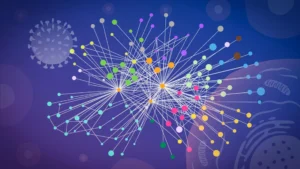
The collaborative team, led by Jan Kosinski of EMBL Hamburg, recognized that these distortions hindered the development of effective antiviral drugs. By integrating specialized cross-linking mass spectrometry (XL-MS) with advanced structural modeling, the researchers successfully "froze" the action within the cell, allowing them to map these fleeting interactions in their native context for the first time.
Supporting Data: Breaking the Barrier of "Broken-Cell" Science
The technical cornerstone of this study was the implementation of a refined version of XL-MS. Developed by Boris Bogdanow and Fan Liu at FMP Berlin, this methodology allows scientists to "cross-link" proteins that are in close proximity while the cell is still intact. This provides a structural snapshot of the interface between viral and host proteins, capturing data that was previously invisible to standard proteomics.
The Power of Predictive Modeling
The researchers paired their experimental findings with a modified version of AlphaFold, the revolutionary protein structure prediction algorithm. While AlphaFold has changed the landscape of biology, its standard use is often limited by a lack of real-world situational data.
"The key advantage of the modified AlphaFold approach is that it allowed us to feed our experimental cross-linking data directly into the structural modeling," explained Kosinski. By providing the model with real-world spatial constraints—essentially telling the algorithm which specific parts of a viral protein were physically touching a host protein—the researchers could generate high-confidence models of virus-host complexes that were previously too complex to predict.
Mapping the Hemagglutinin Pathway
The study’s data highlighted two distinct strategies influenza uses to control the cell. The first involves hemagglutinin, the viral surface protein responsible for anchoring to and entering host cells. The researchers tracked this protein as it moved through the cell’s internal logistics network—the endoplasmic reticulum and Golgi apparatus. Their data revealed that the virus recruits specific human proteins to fold and modify hemagglutinin correctly. Some of these host proteins were previously poorly understood, but the map clarifies their role as vital "assistants" in the viral assembly line.
The Paraspeckle Discovery: A Strategic Dissolution
Perhaps the most striking finding involved the discovery that influenza A actively dissolves "paraspeckles." These are droplet-like structures residing in the cell nucleus that act as regulators for RNA and stress responses.
"What surprised us most was the paraspeckles," said Iuliia Kotova, the study’s first author and a former predoctoral fellow at EMBL. "Watching these tiny organelles in the nucleus dissolve, consistently across every cell line and every flu strain we tested, told us this isn’t a side effect of infection—it might be a strategy."
By destroying these structures, the virus achieves two goals:
- Resource Liberation: The dissolution releases RNA-binding proteins that the virus can then co-opt to support its own replication.
- Defense Neutralization: Because paraspeckles are linked to the cell’s stress response and antiviral gene regulation, their destruction effectively blinds the cell’s internal alarm system, preventing it from mounting a coordinated defense.
Official Responses and Collaborative Synergy
This project was a triumph of interdisciplinary cooperation. The research spanned three major institutions, each contributing specialized technology to solve different parts of the puzzle:
- Charité – Universitätsmedizin Berlin: Provided the specialized cross-linking expertise.
- EMBL Proteomics Core Facility: Conducted the complex glycoproteomics analysis.
- CSSB Advanced Light and Fluorescence Microscopy (ALFM) Facility: Provided the imaging necessary to visualize the internal cellular restructuring.
"Our work provides a new way to study flu-host interactions in their native context and with structural insight," said Jan Kosinski. "The current results are a snapshot of a moment during infection, and it opens the door to studying flu-host interactions across the entire infection cycle."
Boris Bogdanow, now a Junior Research Group Leader at the Institute of Virology at Charité, emphasized the pharmaceutical potential of the work. "This gives us insight into the interface between the virus and the human cell and may, through structural modeling, help identify actionable targets for future pharmaceutical interventions."
Implications: The Future of Pandemic Preparedness
The ability to map molecular contacts within intact, infected cells represents a paradigm shift in virology. By moving away from static, fragmented models, scientists can now observe the "dynamic dance" between pathogen and host.
A Blueprint for Future Viruses
While the current study utilized a laboratory-adapted strain of influenza A, the methodology is designed for scalability. The team believes this approach can be applied to investigate more dangerous, pandemic-potential viruses, such as H5N1. By identifying the precise "hubs" where viral proteins connect to host machinery, researchers can begin to design "pan-viral" drugs—treatments that block the host factors that viruses rely on, rather than just the virus itself.
This is particularly important because, while viruses mutate rapidly to evade vaccines, the host cellular machinery they rely on remains relatively constant. Targeting these conserved interaction sites could provide a robust defense against emerging threats.
Beyond Influenza
The implications of this research extend far beyond the flu. The "mapping in context" approach provides a blueprint for studying how other intracellular pathogens, from SARS-CoV-2 to various hemorrhagic fevers, manipulate the human cell. By combining high-resolution proteomics, structural modeling, and advanced imaging, the scientific community is entering a new era of "systems virology."
In conclusion, the work of Kosinski, Bogdanow, Kotova, and their colleagues has turned a bright light on the shadowy tactics of the influenza virus. By demystifying the molecular sabotage that occurs within our cells, this study has not only deepened our fundamental understanding of viral biology but has also laid the necessary groundwork for the next generation of vaccines and therapeutic interventions. As the threat of future pandemics looms, these molecular maps may prove to be our most valuable tools in the ongoing effort to stay one step ahead of the virus.