










For decades, the medical community has recognized chronic stress as a silent architect of disease, contributing to a litany of ailments ranging from hypertension and insomnia to heart disease and metabolic dysfunction. Yet, despite its profound impact on human longevity, stress has historically remained an elusive, subjective target—often measured by inconsistent patient surveys or cumbersome, fleeting laboratory tests.
That paradigm is now shifting. Researchers from the Johns Hopkins University School of Medicine have unveiled a groundbreaking deep-learning artificial intelligence model capable of identifying the first objective, visual biomarker of chronic stress using standard medical imaging. This innovation, set to be presented at the upcoming annual meeting of the Radiological Society of North America (RSNA), promises to transform how clinicians quantify the cumulative "wear and tear" that stress inflicts on the human body.
The Science of the "Adrenal Gauge"
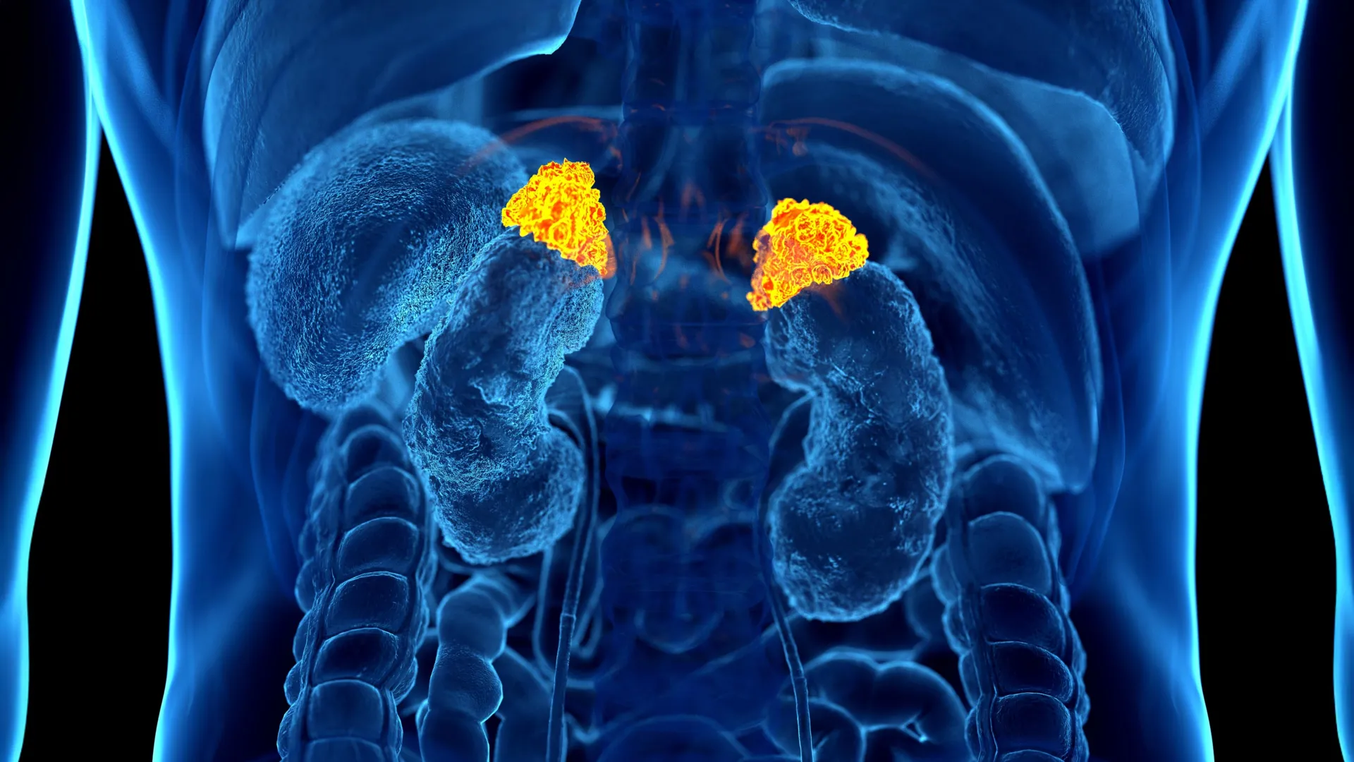
The cornerstone of this research lies in the adrenal glands—small, triangular-shaped organs perched atop the kidneys. These glands are the primary command centers for the body’s stress response, releasing cortisol and other hormones into the bloodstream during periods of strain.
Dr. Elena Ghotbi, a postdoctoral research fellow at Johns Hopkins and the study’s lead author, hypothesized that chronic stress might manifest physically through structural changes in these glands. By training a deep-learning algorithm to analyze thousands of routine chest CT scans, the research team developed a tool to automatically measure the volume of the adrenal glands.
"Our approach leverages widely available imaging data and opens the door to large-scale evaluations of the biological impact of chronic stress," Dr. Ghotbi explained. The resulting metric, dubbed the Adrenal Volume Index (AVI), serves as a long-term, structural barometer of a patient’s stress load, distinct from a single-point-in-time cortisol blood test, which may fluctuate due to temporary factors like a bad night’s sleep or the anxiety of a hospital visit.
A Chronology of Discovery
The journey to this discovery was rooted in the Multi-Ethnic Study of Atherosclerosis (MESA), a massive, longitudinal research endeavor. The path to validating the AVI marker followed a rigorous scientific progression:
- Cohort Selection: The research team tapped into a diverse group of 2,842 participants with a mean age of 69.3. This cohort was uniquely valuable because it provided a rare "triple-threat" of data: high-resolution chest CT scans, comprehensive biological samples, and extensive psychosocial questionnaires.
- Methodological Development: Researchers utilized deep learning to automate the segmentation and measurement of adrenal glands from existing scans. By dividing the total adrenal volume by the square of the patient’s height, they established the AVI, ensuring the measurement was adjusted for individual body size.
- Data Integration: To ensure the AVI was clinically meaningful, the team cross-referenced these measurements with eight daily salivary cortisol samples over two days, as well as an "allostatic load" score—a composite calculation of physiological stress markers including blood pressure, glucose levels, BMI, and white blood cell counts.
- Validation and Correlation: Finally, the researchers compared the AVI scores against the 10-year health outcomes of the participants, specifically looking for correlations with cardiovascular events and heart failure.
Supporting Data: The Link Between Structure and Risk
The data emerging from the study provides a compelling narrative of how psychological stress translates into biological pathology. The research revealed that higher AVI values were consistently linked to:
- Hormonal Dysregulation: Increased overall cortisol exposure and higher peak cortisol levels.
- Systemic Load: Higher allostatic load scores, indicating greater strain across multiple body systems.
- Psychosocial Indicators: Patients who self-reported higher levels of perceived stress and depression consistently demonstrated larger adrenal volumes.
- Cardiovascular Impact: A clear correlation was found between enlarged adrenal glands and an increased left ventricular mass index—a structural change in the heart that is a precursor to heart failure.
Perhaps most critically, the study found that for every 1 cm³/m² increase in AVI, there was a statistically significant increase in the risk of heart failure and death. This establishes the AVI not just as a marker of stress, but as an independent predictor of severe cardiovascular outcomes.
Official Perspectives: A Shift in Clinical Practice
The implications of this study have drawn praise from leaders in epidemiology and radiology, who view it as a milestone in preventative medicine.
"For over three decades, we’ve known that chronic stress can wear down the body across multiple systems," said Dr. Teresa E. Seeman, a professor of epidemiology at UCLA and a co-author of the study. "What makes this work so exciting is that it links a routinely obtained imaging feature with validated biological and psychological measures of stress. It’s a true step forward in operationalizing the cumulative impact of stress on health."
Dr. Shadpour Demehri, a professor of radiology at Johns Hopkins and the senior author of the study, emphasized the clinical accessibility of this tool. "For the first time, we can ‘see’ the long-term burden of stress inside the body, using a scan that patients already get every day in hospitals across the country," he noted.
By utilizing data from existing CT scans, the medical community can potentially integrate stress-risk assessment into standard diagnostic workflows without requiring additional patient visits, radiation exposure, or costly new testing protocols. This is a game-changer for cardiovascular risk stratification, allowing doctors to identify patients who may be at risk for heart failure long before acute symptoms appear.
Implications for Future Health Care
The potential applications of this AI-driven biomarker are vast. In the current healthcare landscape, physicians often rely on patient self-reporting to gauge stress, which is subject to recall bias and social desirability bias. By introducing an objective, physiological measurement, the AVI could allow clinicians to:
- Enhance Preventive Care: Doctors could intervene earlier with lifestyle counseling, stress-reduction therapies, or pharmacological support for patients whose imaging shows elevated adrenal volumes.
- Broaden Diagnostic Scope: While this study focused on cardiovascular risk, the researchers believe the AVI could be applied to a range of other stress-linked conditions, including metabolic syndrome, chronic inflammation, and immune-mediated disorders.
- Standardize Stress Assessment: The integration of AI into routine imaging could effectively turn millions of existing "incidental" CT scans into diagnostic tools for long-term health monitoring.
As the medical field continues to grapple with the "hidden" crises of modern life, the ability to quantify the physiological toll of chronic stress represents a massive leap forward. By making the invisible visible, Dr. Ghotbi and her team at Johns Hopkins have provided a new lens through which we can view—and ultimately manage—the cumulative impact of the modern world on the human body.
As the findings reach the broader community at the RSNA annual meeting, the focus will likely turn toward the clinical implementation of this tool. If adopted widely, the Adrenal Volume Index could redefine our approach to preventative cardiology, moving us away from a reactive model and toward a future where the body’s internal history of stress is used to proactively secure better health outcomes for millions of patients worldwide.