







In a significant breakthrough for regenerative medicine, biomedical engineers at Duke University have unveiled a novel, injectable biomaterial that offers a potential lifeline for victims of ischemic stroke. By transforming the "dead space" left behind by brain injury into a flourishing environment for healing, this technology represents a departure from traditional recovery models. The findings, recently published in the journal Cell Biomaterials, suggest that we may soon be able to do more than simply rehabilitate brain circuits—we may be able to actively guide the brain to rebuild itself.
The Persistence of the Stroke Cavity
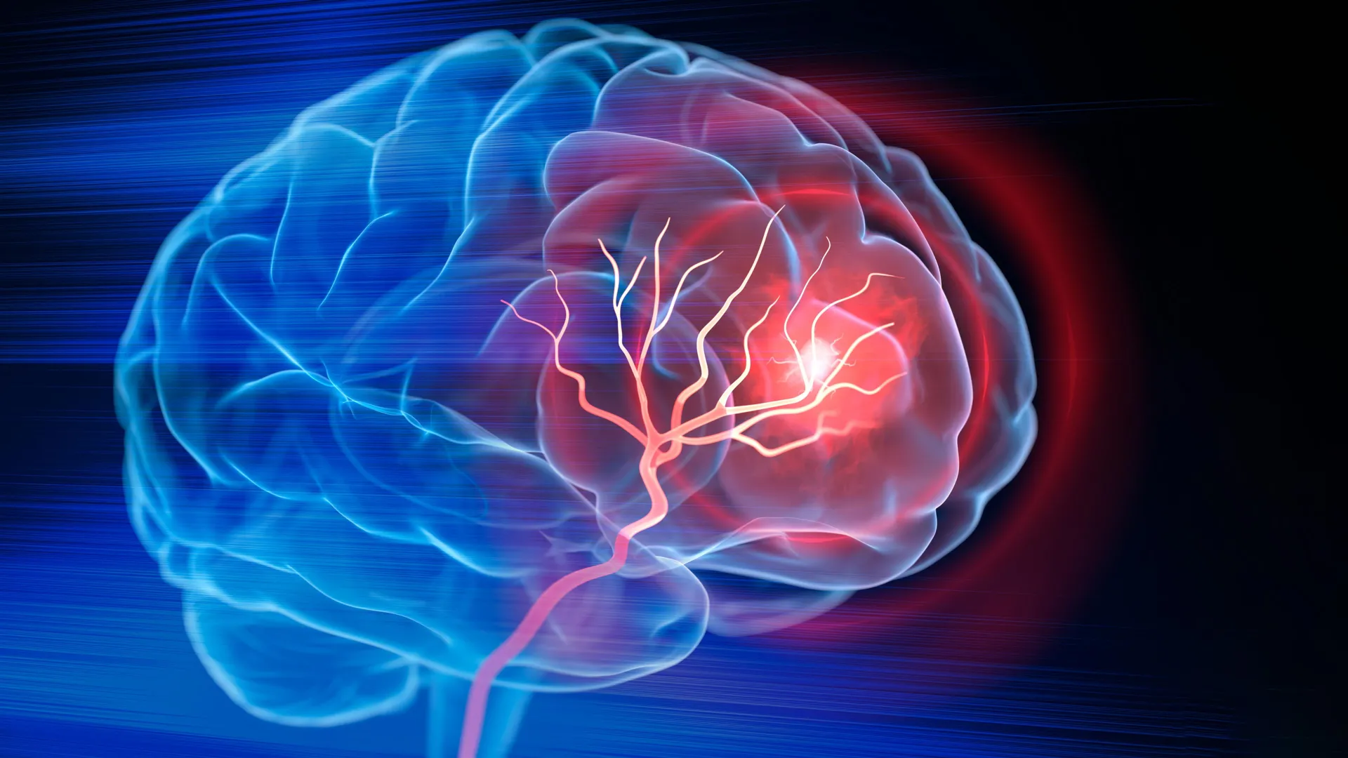
Millions of individuals worldwide suffer from ischemic strokes annually, events triggered when a clot obstructs blood flow, starving brain tissue of oxygen. Modern medicine has made monumental strides in the acute phase of treatment; clot-dissolving drugs and mechanical thrombectomies can successfully restore circulation, salvaging tissue that remains viable.
However, a critical clinical hurdle remains: once brain tissue dies, the damage is often permanent. A severe stroke leaves behind a hollowed-out cavity—a structural void where healthy neural architecture once thrived. Until now, medical intervention for this stage of injury has been largely limited to rehabilitative therapy, which encourages surviving neural circuits to compensate for lost functions. While helpful, this process does not restore the physical or vascular integrity of the damaged area.
"Once brain tissue has been lost, restoring blood flow is no longer enough," explains Tatiana Segura, the Robert Plonsey Distinguished Professor of Biomedical Engineering at Duke University. "Our goal is to engineer the injured space so that immune, vascular, and neural repair processes can begin to work together. We are not simply placing a material into the brain; we are engineering a local environment that can coordinate several parts of the repair response."
Chronology of the Discovery: From Scaffold to Signal
The research team’s journey toward this innovation began with the development of "MAPS"—microporous annealed particle scaffolds. These are comprised of individual hydrogel microparticles that, when injected, self-assemble into a highly porous, sponge-like structure. This open-pore architecture is crucial; it provides a structural framework, or "scaffold," that allows healthy cells to infiltrate and inhabit the void.
Building on the success of these scaffolds, the team sought to address the body’s internal signaling system. They turned their attention to astrocytes—star-shaped glial cells that are essential to brain function and are the first responders to neurological trauma. Astrocytes communicate by releasing extracellular vesicles (EVs), microscopic biological packets containing proteins, lipids, and genetic material.
The researchers theorized that if they could "load" these EVs with specific therapeutic signals and tether them to the MAPS scaffold, they could keep those signals concentrated exactly where they were needed most. This effectively turned the scaffold into a localized, sustained-release therapeutic station.
Supporting Data: Engineering the Immune Response
The experimental results in mouse models were striking. By tethering signaling molecules—specifically IL-4 and C1q—to the scaffold, the team was able to attract and retain specific immune cells within the cavity.
One of the most profound findings involved the recruitment of neutrophils. Traditionally, neutrophils are viewed by clinicians as agents of inflammation that exacerbate secondary damage in the hours and days following a stroke. However, the Duke team observed a more nuanced reality: when these cells were introduced to the scaffold’s optimized environment, they shifted their function from destructive to regenerative.
When the researchers depleted the population of these neutrophils, the results were telling: blood vessel formation plummeted, and the scaffold underwent significantly less remodeling. This confirms that, under the right chemical cues, the immune system can be coaxed into acting as a construction crew for damaged brain tissue.
By the eight-week mark of the study, mice treated with the scaffold demonstrated significant improvement in motor function. In grid-walking tests—a standard assessment for neurological recovery—the treated mice performed with accuracy indistinguishable from healthy control subjects. This recovery was not fleeting; the improvements persisted for the duration of the study, suggesting that the scaffold facilitated a long-term, stable restoration of neural and vascular function.
Official Responses and Scientific Significance
The study’s lead scientist, postdoctoral fellow Shangjing Xin, emphasizes that this research forces a re-evaluation of how we view the inflammatory response post-stroke. "This result changes how we think about neutrophils after stroke," Xin stated. "Their role appears to depend on when they arrive, where they are located, and the signals they receive from their surroundings. Our study demonstrates a potential engineering strategy to recruit and retain these cells at the right time."
The efficacy of the scaffold was further validated by a control test: when the researchers injected the extracellular vesicles alone, without the MAPS scaffold, the repair response was negligible. This underscores the critical importance of the biomaterial’s physical architecture. The scaffold does not merely carry the medicine; it creates the "ecosystem" necessary for the signals to take root.
Clinical Implications: The Road Ahead
While the results are undeniably promising, both Professor Segura and her team acknowledge that the transition from rodent models to human clinical trials is a complex, long-term endeavor.
Key Future Challenges:
- Scalability: Currently, the team uses primary rat astrocytes to source their EVs. For human applications, the researchers are investigating human induced pluripotent stem cell-derived astrocytes. These offer a more sustainable and clinically viable source, with the added benefit of allowing researchers to "program" the vesicles for specific patient needs.
- Safety and Efficacy: While the material successfully integrated in mouse brains, the complexities of the human brain—and the significantly larger volume of tissue damage following a typical stroke—will require extensive safety profiling and rigorous regulatory oversight.
- Translation to Larger Models: Before human trials can be considered, the treatment must prove effective in larger animal models that better mimic the neuroanatomy and recovery timelines of humans.
Rethinking Recovery: A Paradigm Shift
The philosophy behind this research represents a fundamental shift in how we approach the treatment of chronic brain damage. For decades, the consensus in neurology was that the "scars" of a stroke were a permanent reality, and that patients must learn to live with the deficit.
Professor Segura’s vision challenges this notion. "You do not restore an ecosystem simply by containing the initial damage," she notes. "You have to create the conditions that allow life to return. That is how we think about the stroke cavity. The material is not intended to reproduce the brain itself, but to create an environment where the body’s own cells can enter, communicate, and participate in rebuilding vascularized tissue."
By viewing the stroke cavity as a site of potential rather than a site of total loss, this biomaterial offers a new, optimistic trajectory for regenerative neurology. If the success seen in the laboratory can be replicated in clinical settings, this technology could eventually move beyond the stroke ward, potentially offering therapeutic options for other forms of brain injury, including traumatic brain injury (TBI) and neurodegenerative conditions.
As the scientific community watches the progress of this Duke-led initiative, one thing is clear: the future of brain repair may lie not in replacing the organ, but in providing the body with the tools it needs to mend itself. The "scaffold" for a new era of neurology is being built, one microporous particle at a time.