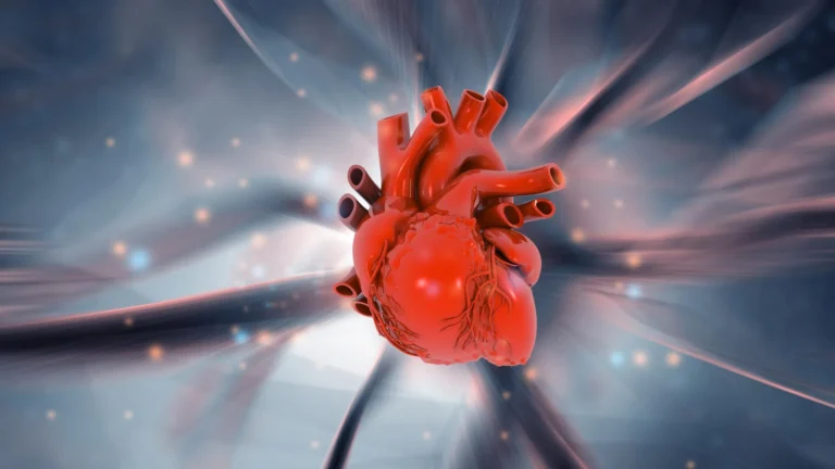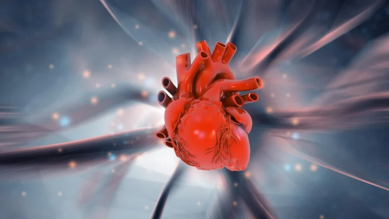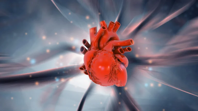









Congenital heart disease (CHD) stands as one of the most pressing challenges in modern pediatrics. Affecting approximately two out of every 100 newborns worldwide, it is the most common category of birth defect, fundamentally altering the lives of millions. Despite its prevalence, the biological "blueprint" that goes awry during fetal development has long remained a complex puzzle for the medical community. However, a landmark study conducted by researchers at the University of Copenhagen has uncovered a previously hidden cellular mechanism—a "molecular antenna"—that may fundamentally shift our understanding of how these defects occur.
The Core Discovery: A Signaling Hub in the Cilium
For decades, scientists have known that embryonic development is a high-stakes orchestration of cellular instructions. Now, researchers have identified a critical communication system on the exterior of cells that acts as a gatekeeper for heart formation.
At the heart of this discovery is the "primary cilium," a microscopic, antenna-like structure that protrudes from the surface of nearly every cell in the human body. While these cilia were once thought to be vestigial, they are now recognized as sophisticated sensory hubs. The team from the University of Copenhagen discovered that a trio of specific proteins—TAK1, TAB2, and PKA-Cα—congregate within these cilia to form a signaling complex. This hub serves as a command center, relaying vital instructions that dictate whether a stem cell will proliferate, migrate, or differentiate into specialized heart muscle tissue.
"We have discovered a new communication system on the exterior of the cell that is crucial for the proper formation of the heart during embryonic development," explains Professor Lars Allan Larsen, an expert in congenital heart disease at the Department of Cellular and Molecular Medicine. "You could say that we have identified an important cog in a highly complex machine."
Chronology of the Investigation: From Patient Data to Laboratory Models
The path to this discovery was not linear; it required an interdisciplinary synthesis of human genetics, developmental biology, and high-tech modeling. The research, recently published in the scientific journal PLOS Biology, followed a rigorous multi-stage investigative process.
Phase 1: Genetic Analysis
The journey began with an extensive analysis of genetic data from thousands of patients diagnosed with congenital heart defects. By comparing the genetic profiles of these patients against healthy control groups, the researchers looked for rare mutations that appeared with statistically significant frequency. This "big data" approach allowed the team to pinpoint specific genes that were likely disrupting the molecular instructions required for cardiac development.
Phase 2: Functional Validation in Zebrafish
Once the candidate genetic mutations were identified, the team needed to prove that these changes were causative rather than correlative. They turned to zebrafish—a standard model in developmental biology due to the rapid growth and transparency of their embryos. Using advanced genetic engineering techniques, the researchers introduced the identified human mutations into the zebrafish. The results were striking: the embryos exhibited clear disturbances in heart formation and a measurable decline in cardiac function, mirroring the defects observed in human patients.
Phase 3: Cellular and Molecular Mechanistic Studies
Finally, the researchers utilized mouse stem cells and laboratory-grown human cell cultures to peer inside the primary cilium. This allowed them to witness the signaling pathway in real-time. By observing what happened when the communication between TAK1, TAB2, and PKA-Cα was disrupted, they were able to map the exact biochemical "breakdown" that leads to structural heart abnormalities.
Supporting Data: The Global Burden of CHD
The gravity of this research is best understood through the scope of the disease it aims to address. According to the World Heart Federation and the Danish Heart Foundation, the statistics regarding congenital heart disease are sobering:
- Global Prevalence: Approximately 2.3 to 2.5 million newborns are affected annually.
- Living Population: As of 2023, an estimated 16 million people are living with the long-term consequences of congenital heart defects.
- Classification: CHD is broadly split into two categories. Non-syndromic CHD involves heart defects occurring in isolation. Syndromic CHD, which was the focus of this study, involves heart defects that appear as part of a wider genetic syndrome, often affecting multiple organ systems simultaneously.
This study provides a unifying explanation for syndromic cases, suggesting that when the "antenna" function is compromised, the failure to process environmental signals leads to a cascade of developmental errors across the body.
Official Responses and Expert Perspective
The implications of this research have been met with enthusiasm by the scientific community. Professor Søren Tvorup Christensen, a lead cell biologist at the Department of Biology, emphasizes that this discovery is not merely about the heart, but about systemic cellular health.
"These proteins act as molecular instructions that tell stem cells when and how to develop into heart muscle cells," says Professor Christensen. "However, genetic alterations can disrupt this communication, causing ‘antenna defects,’ which may lead to congenital heart defects."
The researchers are quick to note the limitations of their study, emphasizing that while the genetic associations are robust and the experimental models in zebrafish provide a clear picture of the mechanism, translating these findings directly into human clinical outcomes remains the next hurdle. "We investigate the mechanism from many different angles and using many different methods," Larsen says. "All of them support what we observe in patients. Therefore, we are reasonably confident that this mechanism also exists in humans."
Broader Implications: Beyond the Heart
Perhaps the most significant takeaway from the study is the realization that the primary cilium’s influence extends far beyond cardiac tissue. Because these cilia are found on nearly every cell type in the human body, the researchers suspect that the mechanism they uncovered may also explain defects in the brain, kidneys, and skeletal system—all of which are often affected in patients with syndromic heart conditions.
"When the ciliary mechanism fails, it typically affects the development of several other organs as well," notes Professor Christensen. "This may explain why some patients with congenital heart disease also have defects and related conditions affecting the brain, kidneys, and skeleton. The mechanism provides a unifying explanation for diseases that we have previously struggled to understand."
The Future of Targeted Medicine
The discovery of this "signaling hub" opens a new door for potential diagnostic and therapeutic interventions. Currently, many rare genetic disorders are categorized by their symptoms rather than their underlying molecular pathology. If scientists can identify patients with specific mutations in the TAK1-TAB2-PKA-Cα pathway early in pregnancy or shortly after birth, it could eventually lead to more personalized medical management.
"Many rare genetic diseases are caused by changes in genes that affect ciliary function, yet the underlying mechanisms have remained poorly understood," Professor Larsen adds. "This new knowledge may eventually make it easier to identify patients early and develop targeted treatments."
Conclusion: A Paradigm Shift in Developmental Biology
The University of Copenhagen study represents a sophisticated marriage of genetics and cell biology, turning a microscopic spotlight on the primary cilium. By defining the role of this cellular antenna, the research team has moved the field closer to a comprehensive "atlas" of human heart development.
While the road from laboratory discovery to clinical application is long, the identification of this specific molecular cog provides researchers with a new target. As the scientific community continues to explore the nuances of the TAK1, TAB2, and PKA-Cα interaction, the hope is that this new understanding will translate into better outcomes for the millions of families worldwide who face the challenges of congenital heart disease every year.
Contributing Researchers:
The study was a collaborative effort involving researchers from the University of Copenhagen, including: Søren Tvorup Christensen, Lars Allan Larsen, Canan Doganli, Oskar Kaaber Thomsen, Daniel A. Baird, Yeasmeen Ali, Menachem V. K. Sarusie, Line Jeanett Jessen, Pauline Munck Truelsen, Johanne Bay Mogensen, Maria Schröder Holm, Lorenzo Buttò, Maria Diamanti, Jindřiška Leischner Fialová, and Lotte Bang Pedersen.