








For millions of people, the term "food allergy" conjures a specific, high-stakes medical scenario: the immediate, life-threatening onset of anaphylaxis, hives, or swelling upon exposure to a trigger. These reactions are typically mediated by Immunoglobulin E (IgE), the antibody responsible for the "classic" allergic response. However, a growing body of clinical evidence suggests that for the Ehlers-Danlos syndrome (EDS) and hypermobility spectrum disorder (HSD) communities, this narrow definition of food allergy is not only incomplete—it is potentially masking a significant source of chronic suffering.
Non-IgE mediated food allergies operate through different immune pathways, often manifesting as delayed, systemic gastrointestinal distress that standard diagnostic tools—like skin prick or IgE blood tests—are fundamentally unable to detect. For patients already navigating the multi-system complexities of connective tissue disorders, these "hidden" allergies represent a critical, yet frequently overlooked, frontier in personalized medicine.
The Main Facts: Defining the Invisible Barrier
Unlike IgE-mediated allergies, which trigger an immediate histamine release, non-IgE mediated food allergies involve a complex interplay of T-cells and other immune components. Because the reaction does not involve the rapid-fire histamine response, symptoms can appear anywhere from several hours to several days after consumption. This latency period makes it notoriously difficult for patients to identify specific dietary triggers without professional guidance.
The primary clinical manifestations of these conditions are gastrointestinal: chronic vomiting, persistent diarrhea, severe abdominal pain, reflux, and malabsorption. These symptoms are frequently dismissed as irritable bowel syndrome (IBS) or simple dyspepsia, leading to years of diagnostic odyssey. The most recognized forms include:
- Food Protein-Induced Allergic Proctocolitis (FPIAP): Typically found in infants, causing inflammation of the colon.
- Food Protein-Induced Enteropathy (FPE): A broader inflammation of the small intestine leading to poor nutrient absorption.
- Food Protein-Induced Enterocolitis Syndrome (FPIES): A severe, delayed reaction that can cause profound vomiting and dehydration, occurring in both children and adults.
For the EDS community, the narrative is further complicated by two specific conditions: Eosinophilic Esophagitis (EoE) and Mast Cell Activation Syndrome (MCAS). These conditions bridge the gap between classic allergy and chronic, non-specific immune activation.
Chronology: A Shift in Medical Understanding
The history of diagnosing food-related immune disorders has been dominated by the IgE paradigm since the early 20th century. However, the last two decades have seen a paradigm shift:
- 2000s: Recognition of EoE as a distinct entity grows, moving away from the assumption that all esophageal inflammation is merely "acid reflux."
- 2010s: Research begins to highlight the high prevalence of gastrointestinal dysfunction in patients with connective tissue disorders, shifting focus toward systemic hypersensitivity.
- 2020–2025: Large-scale studies begin to quantify the "eight-fold risk" of EoE in patients with connective tissue disorders, establishing a formal link between structural protein defects and immune-mediated tissue inflammation.
- 2026 (Present): Clinical guidelines are increasingly advising gastroenterologists to look beyond IgE testing, favoring biopsy and clinical elimination trials for patients with unexplained, chronic gastrointestinal symptoms, particularly those with a history of hypermobility.
Supporting Data: The Biological Link
The correlation between connective tissue disorders and immune dysregulation is no longer considered anecdotal. A landmark study examining over one million patient records confirmed that individuals with EDS and Marfan syndrome face an eight-fold increased risk of developing Eosinophilic Esophagitis compared to the general population.
The TGF-β Connection
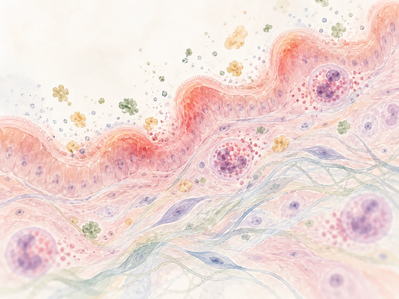
Researchers have identified a potential shared molecular driver: the Transforming Growth Factor-beta (TGF-β) signaling pathway. TGF-β is essential for tissue remodeling and immune regulation. In EDS, where collagen production is faulty, TGF-β pathways are often dysregulated. Because this same protein is a key orchestrator of the inflammatory responses seen in the esophageal lining of EoE patients, scientists believe these two conditions share a common genetic "fault line." This explains why patients with both conditions often experience inflammation that extends beyond the esophagus into the stomach and colon, creating a broader systemic burden.
Official Perspectives and Diagnostic Challenges
The current clinical consensus is that standard allergy testing—specifically skin prick tests and serum IgE panels—is insufficient for diagnosing non-IgE mediated conditions.
The Diagnostic Gold Standard
For EoE, the gold standard remains the endoscopy with biopsy. A physician must directly observe the mucosal lining and count the eosinophil density under a microscope. Relying on blood work to "rule out" allergies in an EDS patient is a clinical error that can delay life-altering dietary interventions.
The Role of MCAS
Mast Cell Activation Syndrome (MCAS) adds another layer of complexity. Mast cells are "first responders" that exist throughout the body, including the gut. When they are hyper-responsive, they release a cascade of mediators that can mimic allergic reactions. Crucially, MCAS can be triggered by non-food factors—stress, temperature, and chemicals—which explains why some patients find that even after eliminating "allergic" foods, they continue to suffer. Management here often involves a combination of H1 and H2 blockers, mast cell stabilizers, and personalized low-histamine diets.
Implications: A New Path for Patient Advocacy
The implications of this research are profound for the EDS community. If you are living with EDS and struggling with persistent GI issues, the following steps are vital for your advocacy:
- Differentiate the Symptoms: Keep a detailed food-and-symptom diary. Because reactions are delayed, identifying a pattern—such as symptoms peaking 24 hours after a specific protein intake—is a powerful tool for your specialist.
- Challenge the "GERD" Diagnosis: If you are being treated for GERD but your symptoms are not resolving, specifically ask your gastroenterologist about the possibility of EoE. Reference the documented eight-fold increased risk associated with connective tissue disorders.
- Seek Specialized Care: Not all gastroenterologists are familiar with the intersection of connective tissue disorders and immune-mediated gut inflammation. Seek out centers that specialize in esophageal disorders or neuro-gastroenterology.
- Advocate for Biopsy: Do not accept a "normal" allergy test as proof that your food-related symptoms are psychosomatic. In non-IgE conditions, the pathology is in the tissue, not the blood.
Moving Forward
The intersection of Ehlers-Danlos syndrome and non-IgE mediated food allergies is a field of active, rapidly evolving research. For the patient, this means the landscape is shifting from "it’s all in your head" or "it’s just your EDS" toward a more rigorous understanding of how our immune systems and connective tissues communicate.
By recognizing that these reactions are distinct, scientifically valid, and highly prevalent in the hypermobile population, patients can move away from passive management and toward proactive, evidence-based care. The path to relief often begins with the simple but revolutionary act of looking beyond the IgE antibody—and listening to the specific, complex needs of the body.
About the Author:
Dr. Jacqueline Moltzau Anderson is a researcher at the University of California specializing in the intersections of systemic immune dysregulation and connective tissue pathology. Her work focuses on bridging the gap between clinical research and patient-centered diagnostic tools.
If you found this information valuable, please share this article to help others in the EDS community find the answers they deserve.