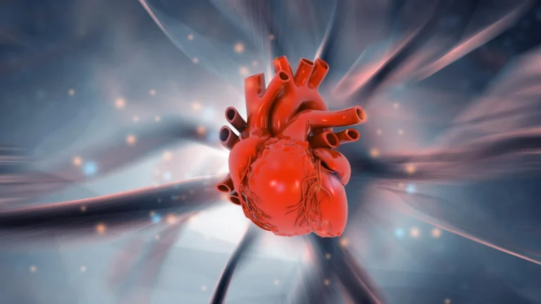












Congenital heart disease (CHD) remains one of the most pressing challenges in pediatric medicine. Affecting approximately two out of every 100 newborns worldwide, these structural abnormalities represent a significant global health burden. Despite the prevalence of the condition, the precise molecular triggers that cause the heart to deviate from its normal developmental path have long remained elusive.
However, a breakthrough study from the University of Copenhagen has shed new light on this complex biological puzzle. By identifying a previously unknown cellular communication system, researchers have pinpointed a crucial "cog" in the embryonic development machine—a discovery that could redefine our understanding of how structural heart defects arise and pave the way for earlier, more targeted medical interventions.
The Microscopic Antenna: Understanding the Primary Cilium
At the heart of this discovery is the primary cilium, a microscopic, antenna-like structure that projects from the surface of nearly every cell in the human body. While long recognized as a sensory organelle, its specific role in cardiac development has been a subject of intense investigation.
The primary cilium functions as a sophisticated biological sensor. It detects chemical signals from the cellular environment—including hormones and growth factors—and translates these external stimuli into internal instructions. These signals dictate fundamental cellular processes, such as whether a cell should proliferate, migrate to a specific location, or undergo programmed cell death.
The University of Copenhagen team, led by Professor Lars Allan Larsen and Professor Søren Tvorup Christensen, discovered that a trio of proteins—TAK1, TAB2, and PKA-Cα—operate as a specialized "signaling hub" within this antenna. This hub acts as a command center, relaying vital instructions to stem cells that dictate their transformation into functional heart muscle cells. When this communication is interrupted, the "antenna" fails, leading to errors in the assembly of the heart’s architecture.
A Chronology of Discovery: From Patient Data to Laboratory Models
The journey to this discovery was a multi-year effort that bridged the gap between clinical genetics and experimental biology. The researchers employed a rigorous, multi-tiered methodology to validate their hypothesis.
Phase 1: Genomic Analysis
The study began with a massive review of genetic data sourced from thousands of patients diagnosed with congenital heart defects. The researchers looked for rare mutations—genetic "typos"—that appeared significantly more often in these patients than in healthy control groups. By isolating these variants, the team identified the specific genes responsible for the malfunctioning signaling hub.
Phase 2: Functional Validation in Zebrafish
Once the candidate genes were identified, the team transitioned to experimental models. Using CRISPR and other genetic engineering techniques, they introduced the identified human mutations into zebrafish embryos. Zebrafish are a gold-standard model for developmental biology due to their transparent embryos and rapid cardiac development. The results were clear: the mutations that disrupted the signaling hub in human cells also caused significant structural heart defects and reduced cardiac function in the zebrafish.
Phase 3: Stem Cell and Laboratory Modeling
Finally, the team utilized mouse stem cells and various laboratory cell cultures to observe the signaling pathways in real-time. This allowed them to map exactly how the loss of communication within the cilium resulted in the failure of stem cells to differentiate into proper heart tissue. By combining human patient data with these controlled experiments, the team created a robust body of evidence that the mechanism they observed in the lab is the same one driving developmental errors in human embryos.
Supporting Data and the Burden of Disease
The significance of this research is underscored by the sheer scale of the global impact of congenital heart disease. According to data from the World Heart Federation and the Danish Heart Foundation, between 2.3 and 2.5 million infants are born with some form of congenital heart defect each year. As of 2023, an estimated 16 million people worldwide are living with the long-term consequences of these conditions.
The study specifically focuses on "syndromic" congenital heart disease. Unlike non-syndromic cases, where the heart defect occurs in isolation, syndromic cases are part of a broader genetic pattern that affects multiple organ systems. Because the primary cilium is found in almost every cell type in the body—from the kidneys to the brain—the researchers noted that the dysfunction of this signaling hub likely explains why many patients with heart defects also suffer from neurological, skeletal, or renal abnormalities.
Official Perspectives: Experts Weigh In
The researchers emphasize that this discovery provides a "unifying explanation" for what were previously considered disparate symptoms in patients with complex genetic syndromes.
"We have identified an important cog in a highly complex machine," says Professor Lars Allan Larsen of the Department of Cellular and Molecular Medicine. "This finding changes our understanding of why some congenital heart defects arise. By investigating the mechanism from many different angles and using many different methods, we are reasonably confident that this mechanism also exists in humans."
Professor Søren Tvorup Christensen of the Department of Biology adds, "These proteins act as molecular instructions that tell stem cells when and how to develop into heart muscle cells. However, genetic alterations can disrupt this communication, causing ‘antenna defects,’ which may lead to congenital heart defects. When the ciliary mechanism fails, it typically affects the development of several other organs as well. This may explain why some patients with congenital heart disease also have defects and related conditions affecting the brain, kidneys, and skeleton."
Implications for Future Medicine
While the study, recently published in the scientific journal PLOS Biology, is a major milestone, the researchers are careful to note that it is only the beginning. Translating these findings into clinical therapies will require further investigation into how these signaling pathways can be modulated or corrected during pregnancy.
Early Detection and Targeted Screening
One of the most immediate potential benefits of this research is the improvement of diagnostic screening. By understanding the specific genes involved in ciliary signaling, doctors may eventually be able to screen expectant parents or newborns more effectively, identifying those at risk of syndromic heart defects before physical symptoms manifest.
New Horizons for Rare Diseases
Beyond cardiology, the implications of this study reach into the broader field of rare genetic disorders. Many rare diseases are known to be caused by mutations that affect ciliary function, yet the biological reasons for their diverse symptoms have remained a mystery. By proving that a single, unified signaling hub can govern the development of multiple organ systems, the University of Copenhagen team has opened the door for new research into other ciliopathies—diseases caused by defective cilia.
Toward Therapeutic Intervention
The long-term goal of such research is to move from simply identifying the causes of birth defects to potentially intervening in their development. While gene therapy is still in its infancy, the ability to pinpoint the precise "molecular instruction" that has gone wrong provides a clear target for future medicinal chemistry and regenerative medicine.
Conclusion: A New Era for Developmental Biology
The discovery that the primary cilium acts as a command center for cardiac development marks a significant shift in developmental biology. By moving beyond the view of the heart as an isolated organ and instead seeing it as a product of highly coordinated cellular communication, the research team has provided a new lens through which to view human development.
As the scientific community continues to explore the nuances of the TAK1, TAB2, and PKA-Cα signaling hub, the hope is that this knowledge will eventually translate into better outcomes for millions of families affected by congenital heart disease. While the path from laboratory bench to clinical bedside is long, this "tiny antenna" has provided a massive leap forward in our quest to understand the building blocks of human life.
Study Contributors:
The research was a collaborative effort involving a large team of scientists from the University of Copenhagen, including Søren Tvorup Christensen, Lars Allan Larsen, Canan Doganli, Oskar Kaaber Thomsen, Daniel A. Baird, Yeasmeen Ali, Menachem V. K. Sarusie, Line Jeanett Jessen, Pauline Munck Truelsen, Johanne Bay Mogensen, Maria Schröder Holm, Lorenzo Buttò, Maria Diamanti, Jindřiška Leischner Fialová, and Lotte Bang Pedersen. The findings were published in the peer-reviewed journal PLOS Biology.