








For three decades, the scientific consensus regarding the development of human central vision was built on a foundation of movement. The prevailing theory suggested that the foveola—the tiny, critical bullseye at the center of the retina—achieved its unique composition of red and green light-detecting cells by physically displacing blue-sensitive cones during fetal development. It was a model of migration: cells were thought to be "born" with a fixed identity and simply moved out of the way to make room for others.
However, a groundbreaking study from Johns Hopkins University, published in the Proceedings of the National Academy of Sciences, has dismantled this long-standing paradigm. By leveraging advanced laboratory-grown retinal organoids, researchers have uncovered a far more dynamic process. It appears that the eye does not merely shuffle its cells; it transforms them. Through a precise, orchestrated interplay of retinoic acid—a derivative of vitamin A—and thyroid hormones, the developing retina undergoes a metamorphosis that fundamentally alters the identity of its photoreceptors.
The Foveola: The Center of Our Visual World
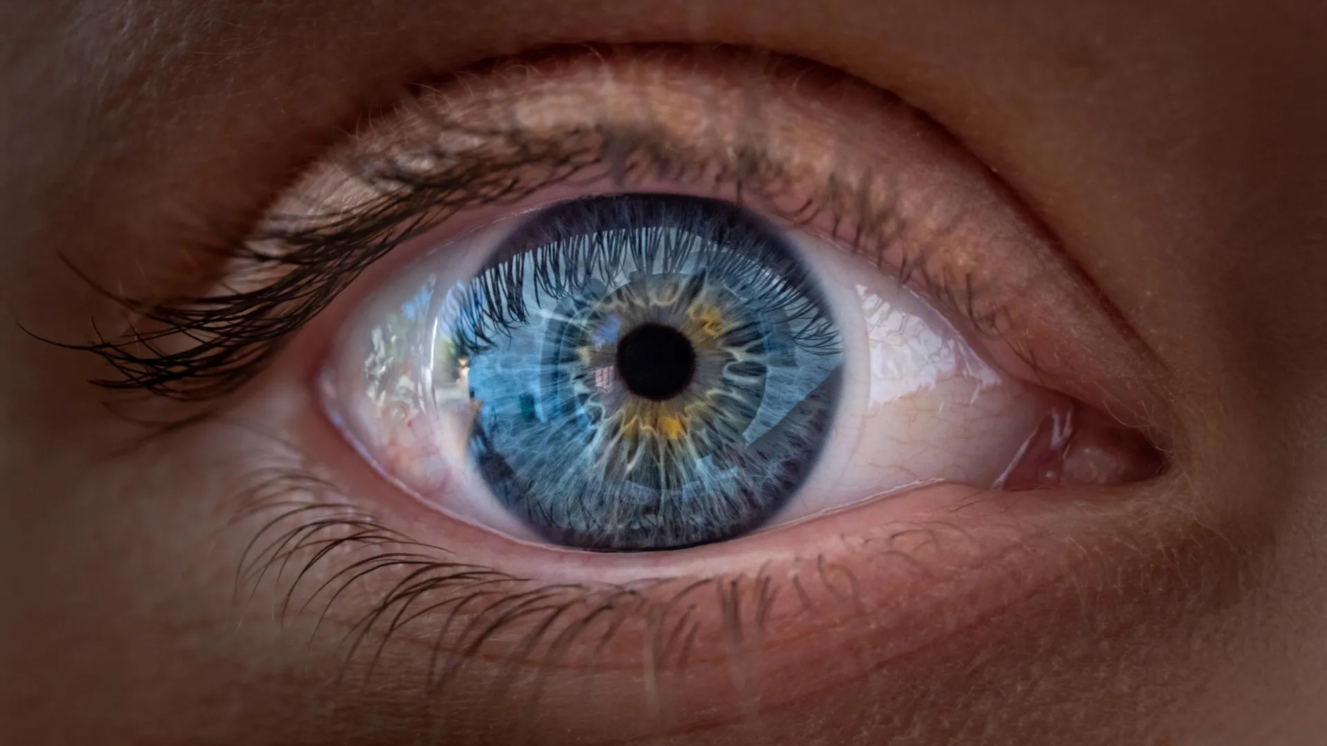
To understand the significance of this discovery, one must appreciate the anatomy of the human eye. The retina, a thin layer of tissue lining the back of the eye, acts as the biological film of a camera, capturing light and converting it into electrical signals for the brain. Within this vast landscape, the foveola is a microscopic but monumental region. Occupying only a tiny fraction of the retinal surface, the foveola is responsible for roughly 50% of human visual perception.
This region is the engine of our sharpest sight. It is where we derive the clarity required to read fine print, thread a needle, or recognize a familiar face across a crowded room. The foveola’s precision is predicated on a specific arrangement of cone cells. While the rest of the retina is populated by a mix of red, green, and blue cones, the foveola is uniquely devoid of blue cones. This "blue-free" zone is the secret behind our ability to resolve high-contrast, high-detail images. For years, scientists puzzled over how this architecture was established, hindered by the fact that common laboratory models—such as mice and fish—lack this specific foveolar organization, making them poor proxies for human retinal development.
A Chronology of Cellular Metamorphosis
The Johns Hopkins research team, led by Associate Professor of Biology Robert J. Johnston Jr., bypassed the limitations of animal models by utilizing "retinal organoids." These are miniature, three-dimensional tissues grown from human fetal cells that successfully mimic the structural and developmental milestones of the actual human eye.
The team’s observations revealed a distinct timeline of events occurring between the 10th and 14th weeks of gestation:
- Weeks 10–12: During the early stages of fetal development, the foveola is not yet a specialized zone. It begins with a nascent population of cone cells, including those sensitive to blue light. Under the traditional model, these cells were thought to be permanent residents waiting to be "pushed out."
- Weeks 12–14: As the retina matures, a critical chemical signal is triggered. Retinoic acid, a derivative of vitamin A, begins to break down in the central region. This degradation acts as a regulatory gatekeeper, limiting the further formation of blue cones in the center of the retina.
- Week 14 and Beyond: Once the "blue-limiting" environment is established by the retinoic acid, thyroid hormones—the body’s metabolic regulators—take the stage. The researchers observed that these hormones act as a biochemical trigger, prompting the existing, "misplaced" blue cones to undergo a genetic reprogramming. These cells shed their blue-light sensitivity and convert into red and green cones, effectively refining the foveola to its mature, high-resolution state.
Challenging a 30-Year-Old Scientific Dogma
The implications of this discovery are profound. By demonstrating that cone cells can undergo an identity shift—transdifferentiating from one type to another—Johnston’s team has challenged the assumption that retinal cells are permanently "fated" early in their development.
"The main model in the field from about 30 years ago was that somehow the few blue cones you get in that region just move out of the way, that these cells decide what they’re going to be, and they remain this type of cell forever," Johnston explained. "We can’t really rule that out entirely, but our data supports a different model. These cells actually convert over time, which is really surprising."
This paradigm shift moves the field away from a mechanical view of retinal formation toward a chemical-signaling model. It suggests that the retina is a highly plastic environment where the final "wiring" of our visual capacity is dictated by the precise timing and availability of specific hormones and vitamins.
Implications for Clinical Vision Restoration
While the study provides a roadmap for how the eye develops in the womb, its most exciting potential lies in the future of clinical medicine. Macular degeneration, a leading cause of blindness in older adults, specifically targets the macula—the area surrounding the foveola. As these cells die, patients lose their central vision, rendering them unable to perform essential daily tasks.
Currently, treatment options for advanced macular degeneration are limited, often focusing on slowing disease progression rather than restoring lost function. The Johns Hopkins findings offer a potential pathway toward regenerative medicine. If scientists can understand the exact chemical signals required to transform stem cells into the specific types of cones found in the foveola, they may eventually be able to "grow" healthy, custom-made photoreceptors in the lab.
Toward "Made-to-Order" Photoreceptors
Katarzyna Hussey, a former doctoral student in Johnston’s laboratory and current molecular biologist at CiRC Biosciences, notes that the organoid technology used in the study is the first step toward a revolutionary new therapeutic approach.
"The goal with using this organoid tech is to eventually make an almost made-to-order population of photoreceptors," Hussey said. "A big avenue of potential is cell replacement therapy to introduce healthy cells that can reintegrate into the eye and potentially restore that lost vision."
However, the researchers remain cautious. Transitioning from a petri dish to a patient’s eye involves monumental hurdles, including ensuring that transplanted cells survive the hostile environment of a diseased retina, successfully integrate into the complex neural circuitry of the eye, and function safely without triggering an immune response.
Setting the Record Straight: Vitamin A Myths
In the wake of the news, the researchers were quick to address a potential public misconception. While the study highlights the critical role of retinoic acid (a vitamin A derivative) in fetal development, this does not imply that adults can improve their vision or reverse macular degeneration by simply increasing their intake of vitamin A supplements.
"This is a key step toward understanding the inner workings of the center of the retina, a critical part of the eye and the first to fail in people with macular degeneration," Johnston stated. "By better understanding this region and developing organoids that mimic its function, we hope to one day grow and transplant these tissues to restore vision."
The study emphasizes that the signaling mechanisms are highly specific to the environment of the developing fetus. In an adult, the environment of the eye is fundamentally different, and the windows for such developmental plasticity have long since closed.
A Future Built on Precision
The work of Johnston and his team represents a masterful application of modern biotechnology to solve an age-old anatomical riddle. By successfully recreating the human retina in the laboratory, they have moved past the limitations of animal studies and gained a high-resolution view of our own biological origins.
As the team continues to refine their organoid models, the focus will shift toward optimization: how to improve the efficiency, safety, and longevity of the cells produced in the lab. While clinical applications are still years, if not decades, away, the path forward is clearer than ever. We now know that the sharpness of our vision is not merely a product of cellular geography, but a complex, timed chemical symphony. By decoding this symphony, science moves one step closer to the ultimate goal: the ability to heal the blind and restore the world to those who have lost the ability to see it in detail.