







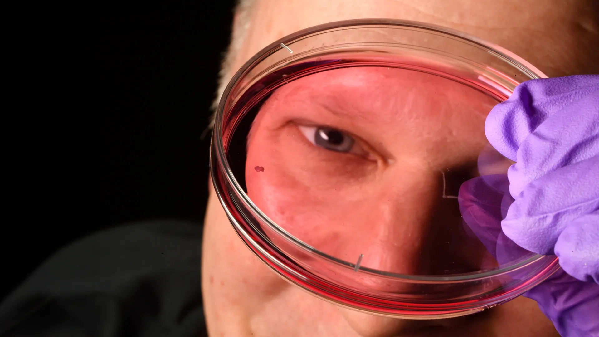
In the intricate theater of human biology, few structures are as sophisticated or as vital as the retina. Within this delicate neural tissue lies the foveola, a microscopic epicenter that grants us the ability to read, recognize faces, and perceive the world in high definition. For decades, the precise developmental choreography that allows the foveola to achieve its singular clarity has remained shrouded in scientific mystery. However, a landmark study from Johns Hopkins University has finally illuminated this process, revealing a biological "remodeling" event that challenges thirty years of established dogma.
By utilizing advanced retinal organoids—lab-grown clusters of fetal cells that mirror the human eye—researchers have discovered that our sharpest vision is not the result of cells migrating away from the center of the eye, but rather a remarkable, hormone-driven identity shift. This discovery, published in the Proceedings of the National Academy of Sciences, marks a pivotal milestone in developmental biology and offers a new roadmap for treating degenerative eye diseases that currently condemn millions to blindness.
The Foveal Paradox: A Decade-Long Scientific Enigma
The human visual system is a masterpiece of evolutionary engineering. Central to this system are the cone photoreceptors: specialized light-sensing cells that provide color vision and high-acuity sight. In the average retina, these cones are distributed in a mosaic of blue, green, and red sensitivities. However, the foveola—the tiny, central pit of the macula—is an anomaly. It is entirely devoid of blue cones, populated instead by a dense, exclusive collection of red and green photoreceptors.
For thirty years, the prevailing scientific consensus suggested a "migration model." Researchers hypothesized that blue cones initially formed in the center of the retina during fetal development and subsequently migrated outward to the periphery to make room for the red and green cells. This theory was largely born of necessity; because the fovea is a uniquely primate feature—and is notably absent in standard laboratory models like mice or zebrafish—the internal mechanics of its formation were largely inaccessible to direct observation.
"The main model in the field from about 30 years ago was that somehow the few blue cones you get in that region just move out of the way," explains Robert J. Johnston Jr., an associate professor of biology at Johns Hopkins University and the lead investigator of the study. "The assumption was that these cells decide their identity early and remain that way forever. Our data suggests a completely different, and far more dynamic, model."
Chronology of Development: A Window into the Fetal Eye
To bypass the limitations of animal models, the Johns Hopkins team turned to retinal organoids. These "mini-retinas" are grown from human fetal cells, allowing scientists to witness the real-time maturation of human tissue in a controlled environment. By tracking these organoids over several months, the team mapped the precise timeline of foveal specialization.
Weeks 10–12: The Initial Blueprint
During the first trimester, the developing foveola begins to take shape. The team observed that in the initial stages, the center of the retina follows the same pattern as the rest of the eye: a mix of blue, red, and green cone precursors begins to emerge. At this stage, the center is not yet specialized; it contains the blue cones that will eventually be phased out.
Week 14: The Great Conversion
By the 14th week of development, a dramatic transformation occurs. The researchers identified a two-stage biological trigger that alters the cellular landscape.
- The Retinoic Acid Brake: First, a molecule derived from vitamin A, known as retinoic acid, is broken down. This degradation is crucial; it effectively halts the production of new blue cones, setting the stage for a population shift.
- The Thyroid Hormone Switch: With the retinoic acid levels reduced, thyroid hormones intervene. These hormones act as a chemical signal that forces the remaining blue-sensitive cells to undergo a genetic reprogramming, converting them into red and green cones.
"First, retinoic acid helps set the pattern. Then, thyroid hormone plays a role in converting the leftover cells," says Johnston. "That’s very important because if you have those blue cones in there, you simply don’t see as well. The foveola requires this specific red-green exclusivity to provide that high-acuity signal to the brain."
Supporting Data and Technical Breakthroughs
The strength of the Johns Hopkins study lies in the unprecedented resolution of their organoid models. Previous research was hampered by the inability to observe the human retina in its embryonic state. By creating tissue that mimics the complex architectural organization of the human eye, the researchers provided the first empirical evidence that cone cells do not necessarily migrate; they transform.
The study utilized advanced genetic sequencing and cellular imaging to track the expression of opsins—the proteins that determine which wavelength of light a cone cell detects. The data showed that the transition from blue-sensitive opsin expression to red/green opsin expression was correlated perfectly with the localized concentration of thyroid hormones within the organoid tissue. This suggests that the foveola is not just a structural pit, but a chemically distinct environment where hormonal gradients dictate the final "wiring" of the visual system.
Implications for Regenerative Medicine
The implications of this discovery extend far beyond developmental biology. Macular degeneration, a leading cause of vision loss in aging populations, specifically targets the center of the retina—the very region this study now understands at a molecular level.
Currently, there are no definitive cures for diseases that destroy photoreceptor cells. However, by identifying the specific molecular "switches"—vitamin A derivatives and thyroid hormones—that govern the identity of cone cells, scientists may eventually be able to instruct stem cells to become the precise type of photoreceptors required for restoration.
Toward "Made-to-Order" Photoreceptors
"The goal with using this organoid tech is to eventually make an almost made-to-order population of photoreceptors," explains Sarah Hussey, a former member of the team and current molecular and cell biologist at CiRC Biosciences.
The vision is to develop cell replacement therapies where healthy, lab-grown photoreceptors are transplanted into the damaged retina. If these cells can be successfully integrated, they could potentially replace the "dead pixels" in the eyes of patients suffering from macular degeneration or glaucoma. While the team cautions that the path from the laboratory to the clinic is long, the discovery provides the "how-to" manual for building a functional retina from the ground up.
Official Responses and Future Directions
The scientific community has reacted with significant interest to the Johns Hopkins findings. By overturning a thirty-year-old assumption, the study highlights the necessity of human-derived tissue models in understanding human-specific anatomy.
"These are very long-term experiments," notes Hussey. "Of course, we would need to do rigorous optimizations for safety and efficacy studies before moving into the clinic. We have to ensure that these transplanted cells don’t just survive, but integrate properly into the existing neural circuitry. It’s a viable journey, but it requires extreme precision."
The Johnston lab is already working to refine their organoids to better mimic the functional complexity of the adult human retina. Future studies will focus on whether these hormonal triggers can be used to "reprogram" damaged or dormant cells within living subjects, potentially offering a non-surgical avenue for vision restoration.
Conclusion: A New Horizon for Ophthalmology
The discovery of the mechanism behind foveal formation is a testament to the power of modern organoid technology. By peering into the development of the human eye, Johns Hopkins researchers have moved past the outdated "migration" theory and identified the sophisticated chemical signaling that makes human color vision possible.
As we stand on the precipice of a new era in regenerative medicine, this work provides more than just an answer to an old question; it provides a blueprint for the future of sight. Whether through cell transplantation or targeted hormonal therapy, the ability to control the identity and development of our most vital sensory cells represents one of the most promising frontiers in modern medicine. For those currently facing the darkness of macular degeneration, this research offers a flicker of hope—a scientific roadmap that may one day turn the tide on vision loss.