










In a landmark advancement for oncology, researchers at Weill Cornell Medicine and the Cornell Duffield College of Engineering have developed a pioneering class of silica-based nanoparticles capable of executing a "one-two punch" against aggressive prostate cancer. By simultaneously triggering internal cellular destruction and reawakening the body’s dormant immune response, these ultrasmall particles have achieved unprecedented success in preclinical trials, signaling a potential shift in how medicine approaches the treatment of solid tumors.
The study, published in the June 15 issue of the journal Cancer Research, details how these engineered particles—known as "Cornell Prime dots" or C’ dots—not only induce tumor cell death but also transform the immunosuppressive environment surrounding the cancer into an immune-active landscape. For prostate cancer, a disease where durable responses to immunotherapy have historically proven elusive, this dual-mechanism approach could represent the dawn of a new clinical paradigm.
The Core Facts: A New Weapon Against Cancer
At the heart of this innovation is a material as unassuming as it is powerful: amorphous silica. This form of silicon dioxide is a naturally occurring compound, found in everything from the fossilized remains of microscopic organisms to common dietary staples like leafy greens and cereal grains.
The C’ dots are engineered as ultrasmall, fluorescent core-shell structures. Initially conceptualized for high-precision medical imaging and image-guided surgery, these particles have already passed the rigorous safety hurdles required for late-stage human clinical trials. However, it was only recently that the collaborative research team discovered the particles’ inherent, selective lethality toward cancer cells.
The nanoparticles are designed to seek out and attach to Prostate-Specific Membrane Antigen (PSMA), a protein that acts as a beacon on the surface of prostate tumor cells. Once docked, the particles initiate a cascade of events that leads to the systematic dismantling of the tumor, all while sparing the healthy, surrounding tissues.
Chronology of Discovery: From Imaging to Intervention
The evolution of C’ dots is a testament to the power of interdisciplinary collaboration, spanning over a decade of work between the laboratory of Dr. Michelle Bradbury, an expert in radiology and molecular imaging, and the laboratory of Dr. Ulrich Wiesner, a pioneer in materials science and engineering.
Phase 1: The Imaging Origins
The C’ dots were first developed to address a critical need in surgery: how to see the "invisible." By providing high-contrast imaging, the dots allowed surgeons to distinguish tumor margins from healthy tissue with pinpoint accuracy. Their success in clinical imaging trials laid the essential groundwork for their current biological application.
Phase 2: The Accidental Breakthrough
As the team studied the biocompatibility and behavior of the particles in complex biological environments, they began to notice a pattern. The particles were not merely "passive observers" in the body. In the presence of tumor cells, they demonstrated a selective toxicity that did not extend to healthy cells. This led to the hypothesis that the particles could be re-engineered from diagnostic tools into therapeutic agents.
Phase 3: Mechanism Validation
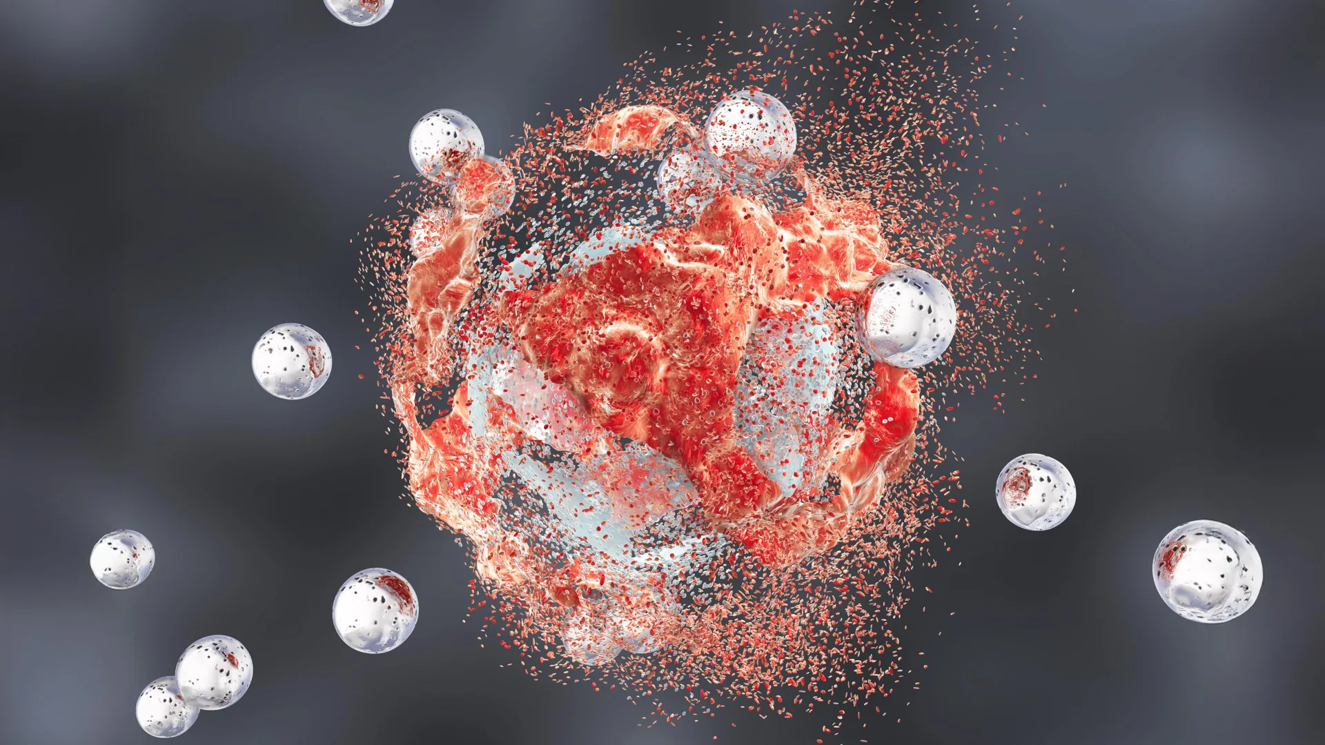
The researchers pivoted their efforts to understand how these particles killed cancer. The resulting experiments identified "ferroptosis"—a specialized, iron-dependent form of cell death—as the primary executioner. The study confirmed that the particles could effectively traffic iron ions into the tumor cell, triggering oxidative stress that destroys the cell’s fatty membranes, ultimately causing the tumor to collapse from within.
Phase 4: Immune Remodeling
The final stage of the current research involved observing the systemic effects of the treatment. The team realized that the destruction of tumor cells was just the beginning. By disrupting the metabolic processes within the tumor microenvironment, the C’ dots effectively "woke up" T cells and macrophages, turning the tumor from a "cold" (immune-resistant) site into a "hot" (immune-active) site that the body’s own defenses could recognize and attack.
Supporting Data: The Power of Combination Therapy
The efficacy of the C’ dots was tested in mouse models harboring aggressive, treatment-resistant prostate cancer. The findings were not merely encouraging; they were transformative.
Survival Statistics
- Monotherapy: When used alone, both the C’ dots and standard immunotherapy drugs showed only a modest improvement in survival rates compared to the control group.
- Synergistic Impact: When the C’ dots were combined with immune checkpoint blockade therapy, the results shifted dramatically. Four out of ten mice achieved complete or near-complete remission, showing no sign of tumor growth and surviving indefinitely.
- Triple Therapy: The researchers added a third component—a CSF-1R blockade designed to target tumor-associated macrophages. This combination pushed the success rate to five out of ten mice, a significant milestone for a disease known for its aggressive resistance to conventional therapy.
Beyond the survival data, the team reported an absence of systemic toxicity. Despite the particles accumulating briefly in organs like the spleen, the targeted nature of the PSMA-binding ensured that the destructive oxidation was confined strictly to the cancerous tissues.
Official Responses and Scientific Perspective
The research team, which includes a wide array of scientists and oncologists from Weill Cornell Medicine and NewYork-Presbyterian, has expressed cautious but profound optimism regarding these findings.
Dr. Michelle Bradbury, senior author and director of the Molecular Imaging Innovations Institute, emphasized the potential for a paradigm shift. "We’re very encouraged by these results; a treatment that directly induces tumor-cell death while transforming the immune microenvironment, as this does, would represent a new clinical paradigm," she stated.
Dr. Ulrich Wiesner, who co-led the study, expressed wonder at the simplicity and efficacy of the material. "It seems unreal—how is it possible that rather than a single pathway we see all these effects happening simultaneously and only in tumors and not in healthy tissues?" he remarked. "I have to wonder whether ultrasmall silica’s very early and ubiquitous presence in the environment and foods like leafy greens or cereal grains has given it a connection to biology that we’re only beginning to glimpse."
Dr. Jedd Wolchok, a co-author and oncologist, highlighted the clinical necessity of this discovery. "One of the most intriguing aspects of this work is the convergence of direct tumor cell killing with broad immune remodeling," Dr. Wolchok said. "By creating conditions that support a more effective antitumor immune response, these particles may help unlock the full potential of immunotherapy in prostate cancer, where durable responses have historically been difficult to achieve."
Implications for the Future of Oncology
The implications of this study extend far beyond prostate cancer. The researchers believe that the "dual-action" mechanism of C’ dots—the ability to kill cells directly while simultaneously re-engineering the immune environment—could be applied to a variety of solid tumors.
Overcoming Resistance
One of the greatest challenges in modern oncology is "immune resistance," where tumors develop barriers that prevent T cells from attacking them. By effectively "heating up" these cold tumors, C’ dots could revitalize existing immunotherapy drugs that have failed in previous trials.
The Path to Human Clinical Trials
With the preclinical work yielding robust results, the primary objective of the collaborative team is now to transition the technology into human clinical trials. The fact that the base technology (the C’ dot) has already navigated the regulatory hurdles for imaging provides a distinct advantage, potentially accelerating the timeline for safety and efficacy testing in human patients.
Collaborative Perseverance
The researchers were quick to credit the immense team effort required to reach this stage. Co-first authors Dr. Nabil Siddiqui, Dr. Li Zhang, and Dr. Gabriel DeLeon, alongside graduate students Nada Naguib and Rachel Lee, were instrumental in the mechanistic and translational studies. As Dr. Bradbury noted, this project stands as a benchmark for what can be achieved when materials scientists and clinicians work in lockstep to solve the most complex problems in human health.
While much work remains before these nanoparticles reach the pharmacy shelf, the research offers a glimpse into a future where cancer therapy is not just about poisoning the tumor, but about empowering the body to destroy it from within. Supported by funding from the Department of Defense, the National Cancer Institute, and the Parker Institute for Cancer Immunotherapy, the Cornell team remains at the vanguard of this new, silica-driven frontier.