








In a breakthrough that fundamentally alters our understanding of human ocular development, researchers at Johns Hopkins University have identified the precise biological mechanism that grants humans the gift of sharp, central vision. By leveraging advanced organoid technology, the team has mapped a carefully timed, complex interplay between vitamin A derivatives and thyroid hormones—a discovery that not only settles a decades-old scientific debate but also lays the groundwork for regenerative therapies aimed at curing currently untreatable forms of blindness.
The findings, recently published in the Proceedings of the National Academy of Sciences, describe how the human retina—the light-sensitive tissue at the back of the eye—sculpts the foveola, the tiny, highly specialized region responsible for our most acute visual acuity. This discovery challenges the long-standing "migration" theory of retinal development and provides a blueprint that could one day be used to grow replacement tissue for patients suffering from macular degeneration and glaucoma.
The Foveola: The Epicenter of Human Vision
To understand the magnitude of this discovery, one must first understand the architecture of the human eye. The foveola is a microscopic region at the very center of the retina. Despite occupying only a fraction of the total retinal surface, the foveola is responsible for approximately half of all human visual perception. It is the seat of high-resolution vision, the mechanism that allows us to read text, recognize faces, and perceive the fine details of the world.
The human retina is populated by millions of photoreceptor cells known as cones. These cells are responsible for daytime and color vision, categorized by their sensitivity to specific wavelengths of light: red, green, and blue. While the broader retina features a mosaic of all three cone types, the foveola is unique; it is almost entirely devoid of blue-sensitive cones, housing instead a highly dense concentration of red and green cones. This specific arrangement is what allows for the sharpness we experience in our direct line of sight.
Chronology of Development: From Fetal Cells to Specialized Sight
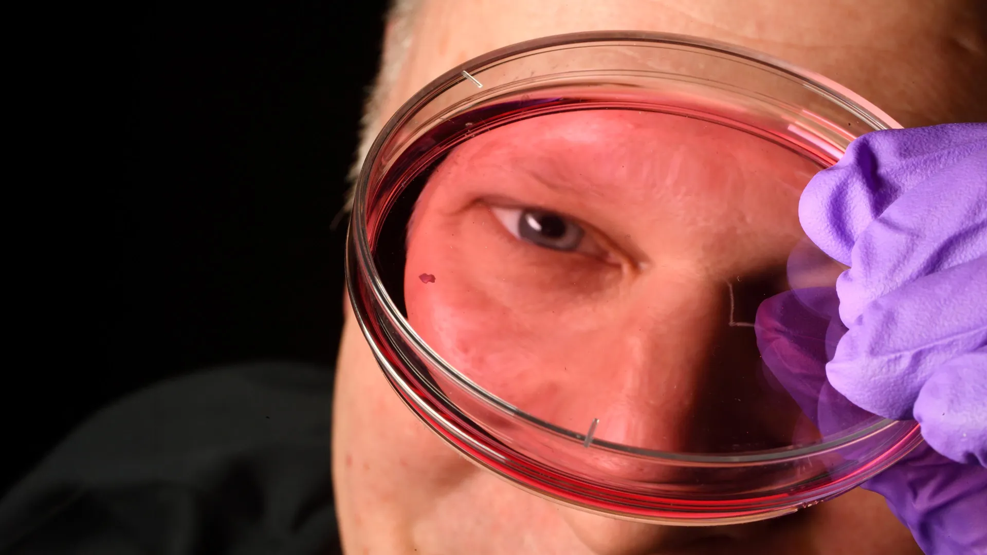
The research team, led by Robert J. Johnston Jr., an associate professor of biology at Johns Hopkins, utilized retinal organoids—miniature, lab-grown clusters of tissue derived from human fetal stem cells. By observing these organoids over several months, the researchers were able to track the cellular "choreography" of the developing eye in real-time.
The 10–12 Week Window
The study pinpointed the critical period of development between the 10th and 12th weeks of gestation. During this initial stage, the developing foveola begins to populate with a small number of blue-sensitive cones. For decades, the prevailing consensus was that these blue cones were a permanent fixture of that region, and that their absence in the adult foveola was due to the cells physically migrating away from the center.
The 14-Week Transformation
By the 14th week, the researchers observed a startling shift: the blue cones were no longer present in the center, but they had not moved. Instead, they had undergone a radical identity shift. Through a two-step molecular process, these cells transitioned into red and green cones.
The first step involves the breakdown of retinoic acid, a molecule derived from vitamin A. The reduction of retinoic acid limits the production of new blue-sensitive cones. Once this stage is set, the second, more dramatic step occurs: thyroid hormones enter the cellular environment and force the remaining blue-sensitive cones to convert their chemical machinery, effectively "reprogramming" themselves into red and green cones.
Challenging the 30-Year Dogma
For over three decades, the primary model in the field of ophthalmology held that cone cells were "fixed" in their identity early in development. According to this traditional theory, if a cell began its life as a blue cone, it remained a blue cone forever. If a region of the retina lacked blue cones, it was assumed that those cells had simply relocated to the periphery of the eye.
"The main model in the field from about 30 years ago was that somehow the few blue cones you get in that region just move out of the way," Johnston explained. "Our data supports a different model. These cells actually convert over time, which is really surprising."
This paradigm shift is significant because it suggests that the retina is far more plastic and malleable than previously believed. By proving that photoreceptors can change their spectral identity, Johnston’s team has opened a door to the possibility of manipulating these cells in clinical settings.
Supporting Data and Methodology
The study relied on high-resolution imaging and molecular analysis of retinal organoids, which provide a reliable proxy for human development. Because common research models—such as mice and fish—do not possess the same foveal structure as humans, studying this process in live animal models has been historically impossible.
The organoids successfully mimicked the human retina’s structural development, allowing the team to document the specific gene expression changes associated with the thyroid hormone conversion. By isolating the role of the thyroid receptor, the researchers confirmed that the absence of these hormones prevents the conversion, leaving behind an abundance of blue cones—a finding that mirrors certain rare color-vision deficiencies.
Official Responses and Implications for Medicine
The implications of this research extend far beyond the fundamental understanding of biology. For millions of people worldwide, the degradation of the fovea is a life-altering experience. Diseases such as age-related macular degeneration (AMD) specifically target the center of the retina, leading to a loss of central vision that renders tasks like driving, reading, and recognizing faces impossible.
A Path Toward Regenerative Medicine
"This is a key step toward understanding the inner workings of the center of the retina, a critical part of the eye and the first to fail in people with macular degeneration," Johnston noted.
The goal for the Johns Hopkins team is now to refine these organoids to serve as a manufacturing platform for healthy photoreceptor cells. By mastering the hormonal "instructions" that dictate cell identity, scientists hope to eventually cultivate specific types of cone cells in the laboratory that can be transplanted into the eyes of patients.
"The goal with using this organoid tech is to eventually make an almost made-to-order population of photoreceptors," said Sarah Hussey, a former member of the research team and current molecular and cell biologist at CiRC Biosciences in Chicago. "A big avenue of potential is cell replacement therapy to introduce healthy cells that can reintegrate into the eye and potentially restore that lost vision."
Future Horizons: The Long Road to the Clinic
While the results are undeniably historic, the researchers are careful to temper expectations regarding immediate clinical application. Restoring vision through cell replacement is an incredibly complex engineering feat. The transplanted cells must not only be healthy but must also physically integrate into the existing neural network of the retina and successfully transmit signals to the brain via the optic nerve.
"These are very long-term experiments," Hussey added. "Of course, we’d need to do optimizations for safety and efficacy studies prior to moving into the clinic. But it’s a viable journey."
The next phase of the research will focus on the longevity and stability of these lab-grown cells. The team plans to investigate how these photoreceptors react to different environments and how they can be scaled up for potential therapeutic use.
A New Era of Visual Science
The discovery of the thyroid-driven conversion of cone cells represents a triumph of modern developmental biology. By peering into the microscopic, dark environment of the fetal eye, the researchers at Johns Hopkins have illuminated the process by which we see the world. As these findings are peer-reviewed and integrated into the broader field of vision science, they provide more than just an answer to a 30-year-old mystery; they offer a flicker of hope to those for whom the world has gone dark. The blueprint for human vision is no longer a locked vault; it is a map, and for the first time, researchers know which pathways to follow.