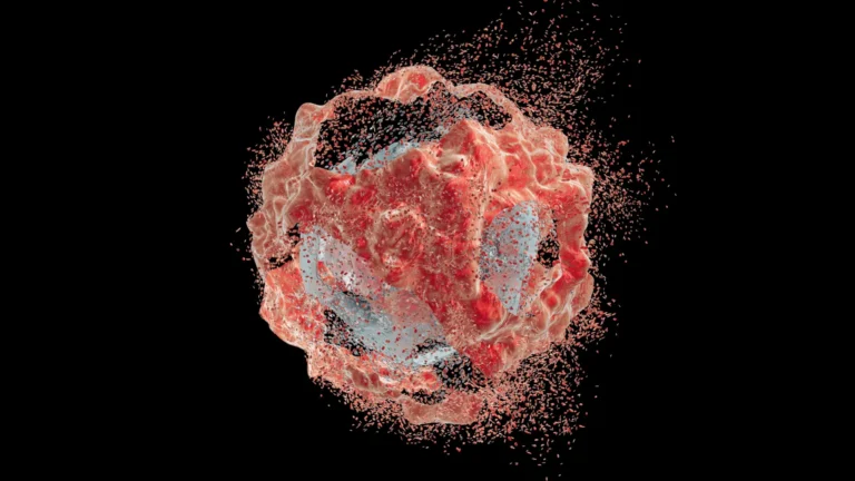









Every year, seasonal influenza strikes millions, resulting in up to 650,000 deaths worldwide. Beyond its annual toll, the virus remains a perennial candidate for global pandemics, with the 1918 Spanish Flu serving as a grim reminder of its lethality. For decades, virologists have sought to understand exactly how the influenza A virus (IAV) transforms a healthy human cell into a sophisticated factory for its own replication.
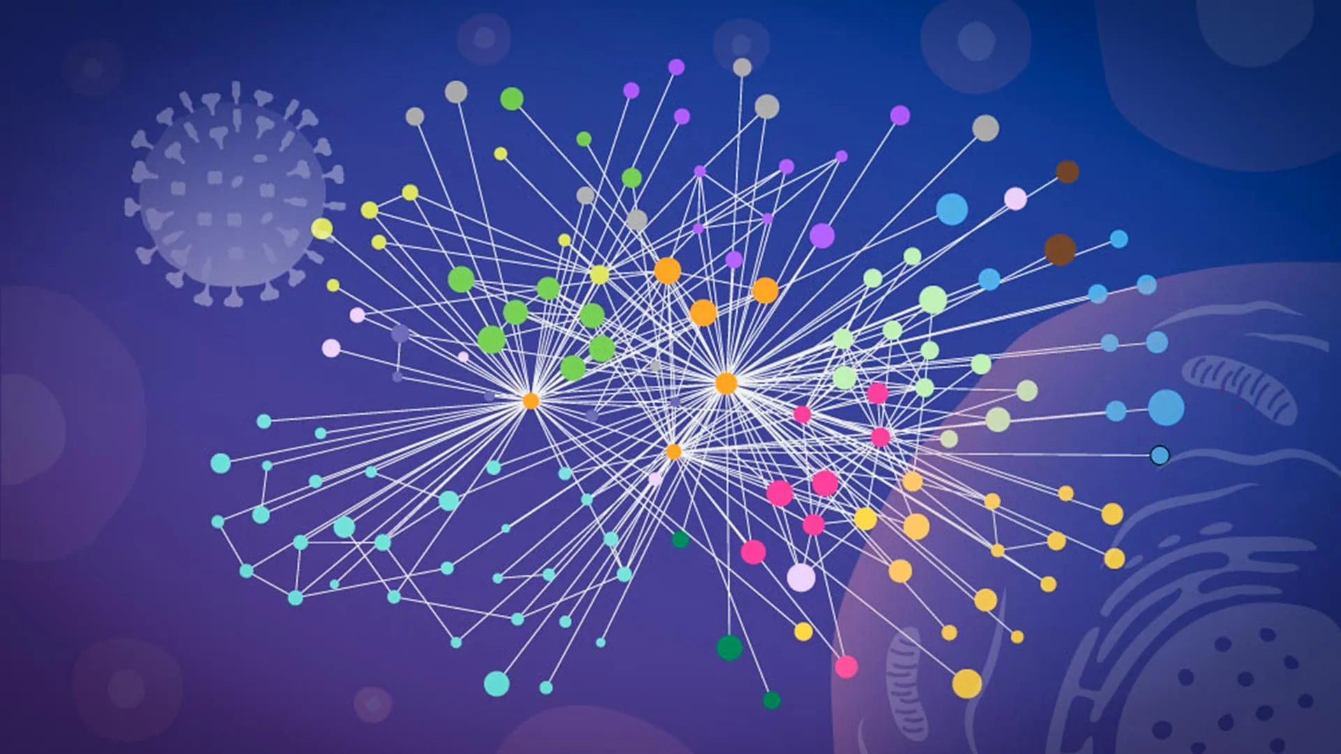
Now, a groundbreaking study led by researchers at EMBL Hamburg, in collaboration with the Leibniz Research Institute for Molecular Pharmacology (FMP) and Charité – Universitätsmedizin Berlin, has produced the most detailed map to date of the virus-host interface. By capturing molecular interactions directly inside intact cells, the team has revealed how influenza systematically rewires cellular machinery, offering a blueprint that could fundamentally shift the development of future antiviral drugs and vaccines.
The Challenge of the "Black Box"
To understand the significance of this research, one must first understand the limitations of traditional virology. For years, scientists studying protein-protein interactions—the primary language of cellular life—relied on "breaking" the cell. To see which proteins were touching, researchers would rupture the cell membrane and isolate its contents.
However, this destruction comes at a cost. When a cell is lysed, its highly organized internal compartments are obliterated. Proteins that were once separated by spatial barriers suddenly collide in the laboratory test tube, creating "false positives" that do not exist in a living system. Conversely, the fragile, transient, or location-specific interactions that are essential to the viral lifecycle are often lost or disrupted during the extraction process.
"The current results are a snapshot of a moment during infection, but they open the door to studying flu-host interactions across the entire infection cycle," said Jan Kosinski, Group Leader at EMBL Hamburg and the Centre for Structural Systems Biology (CSSB). By moving beyond the test tube, the research team has effectively moved from looking at blurry, reconstructed snapshots to viewing high-definition "live-action" footage of the infection process.
A Chronology of Discovery: From XL-MS to AlphaFold
The breakthrough was made possible by an interdisciplinary approach that fused advanced biochemistry with cutting-edge artificial intelligence.
Step 1: Specialized Cross-Linking Mass Spectrometry (XL-MS)
The team turned to a technique known as cross-linking mass spectrometry (XL-MS). While XL-MS has long been used to map protein contacts, the team at FMP Berlin, led by Boris Bogdanow and Fan Liu, developed a specialized version of the technique optimized specifically for virus-infected cells. This allows scientists to "freeze" proteins in their tracks while they are still inside the cell, using chemical cross-linkers to lock interacting partners together before the cell is processed.
Step 2: Integrating Computational Modeling
Once the "frozen" interactions were captured, the researchers faced the challenge of interpreting the structural data. They employed a modified version of AlphaFold, the Nobel Prize-winning protein structure prediction algorithm. By feeding their experimental cross-linking data directly into the AlphaFold model, the researchers were able to simulate not just which proteins were interacting, but how they were physically positioned relative to one another. This was critical for visualizing the virus-host complexes, which are notoriously difficult to predict using purely computational methods.
Step 3: Validation and Analysis
The final phase involved rigorous validation. Using microscopy imaging at the CSSB’s Advanced Light and Fluorescence Microscopy (ALFM) Facility and glycoproteomics at the EMBL Proteomics Core Facility, the team confirmed their findings, ensuring that the structural models aligned with the biological reality observed under the lens.
Two Faces of Viral Hijacking
The study, published in Nature Microbiology, highlighted two specific, high-stakes strategies influenza A employs to hijack host cells.
1. The Hemagglutinin Supply Chain
The influenza virus relies on a surface protein called hemagglutinin to attach to and enter human cells. Once inside, the virus must navigate the host’s complex protein-processing network—the "factory floor" of the cell—where proteins are folded and modified before being shipped to their final destinations. The research revealed that the virus recruits specific human proteins to act as chaperones for hemagglutinin. These host proteins ensure the viral protein is folded correctly, effectively forcing the cell to provide quality control for the virus’s own components. Many of these host proteins were previously poorly understood, marking them as potential new targets for therapeutic intervention.
2. The Dissolution of Paraspeckles
Perhaps the most surprising finding concerned "paraspeckles"—small, droplet-like organelles nestled within the cell nucleus. These structures are involved in cellular stress responses and the regulation of gene expression. The researchers observed that influenza A infection causes these paraspeckles to systematically dissolve.
"Watching these tiny organelles in the nucleus dissolve, consistently across every cell line and every flu strain we tested, told us this isn’t a side effect of infection—it might be a strategy," explained Iuliia Kotova, the study’s first author. By breaking down these structures, the virus releases RNA-binding proteins that it likely repurposes to support its own replication. Furthermore, by dismantling these stress-response centers, the virus may be actively disabling the cell’s internal alarm system, preventing the host from mounting an effective antiviral defense.
Implications for Future Medicine
The ability to map these interactions within the native cellular environment is a game-changer for pharmaceutical research. Current antivirals often target the virus directly, which can lead to the rapid emergence of drug-resistant strains. By identifying the specific host proteins that influenza relies on, researchers can potentially develop "host-targeted" therapies. Because these human proteins are essential for the virus but less prone to rapid mutation, they represent a more stable target for long-term drug development.
"This gives us insight into the interface between the virus and the human cell and may, through structural modeling, help identify actionable targets for future pharmaceutical interventions," noted Boris Bogdanow, now a Junior Research Group Leader at the Institute of Virology at Charité – Universitätsmedizin Berlin.
A Collaborative Blueprint for Pandemic Preparedness
The success of this study was rooted in a unique cross-institutional synergy. The project utilized the expertise of the EMBL compute cluster for structural modeling, the FMP for mass spectrometry, and the CSSB for advanced microscopy. This collaborative model demonstrates that solving the mysteries of complex pathogens requires more than just biological expertise—it requires the integration of physics, chemistry, data science, and engineering.
While the study focused on a laboratory-adapted strain of influenza, the methodology is designed for scalability. The team believes this approach can be applied to investigate other high-pathogenicity viruses, including avian influenza strains like H5N1. By uncovering the "hidden networks" that support viral multiplication, scientists are now better equipped to anticipate how these viruses adapt to human cells.
As Jan Kosinski concluded, the "mapping in context" approach is likely to become a standard tool in the virologist’s arsenal. Whether investigating seasonal flu or preparing for the next potential pandemic, the ability to observe the virus in its "native context" represents a significant leap forward in our quest to outsmart one of humanity’s oldest and most persistent adversaries. Through the lens of this research, the virus is no longer a black box—it is a map, and for the first time, we can see exactly where the virus is heading, and how we might stop it in its tracks.