









Introduction: The Silent Invaders
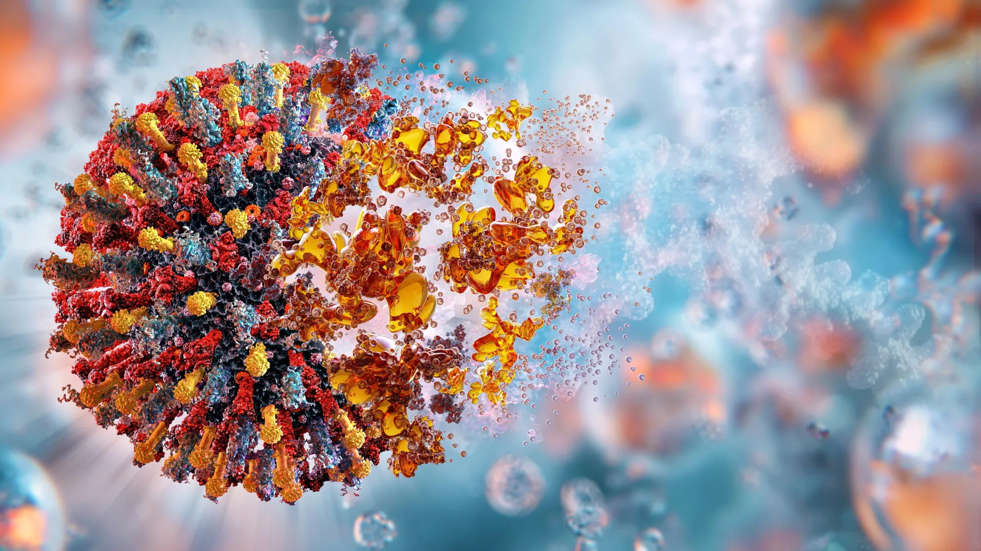
Enteroviruses are among the most persistent and versatile pathogens known to medicine. From the debilitating paralysis historically associated with polio to the modern-day concerns of myocarditis, encephalitis, and the ubiquitous common cold, these viruses have long posed a significant challenge to global health. Despite their profound impact, the fundamental mechanics of how these microscopic agents seize control of human cellular machinery have remained shrouded in mystery.
Now, a pivotal discovery by researchers at the University of Maryland, Baltimore County (UMBC) has shed light on this biological "black box." Published in the journal Nature Communications, the study details exactly how enteroviruses hijack a host cell’s resources to replicate their genetic material. By mapping the precise molecular interactions between viral RNA and specialized proteins, the team has not only solved a long-standing scientific puzzle but has also illuminated a promising new pathway for the development of broad-spectrum antiviral medications.
The Mechanics of Hijacking: A Scientific Chronology
The research, spearheaded by Deepak Koirala, an associate professor of chemistry and biochemistry at UMBC, alongside recent Ph.D. graduate Naba Krishna Das, represents the culmination of years of rigorous investigation into the structural biology of RNA viruses.
The Cloverleaf Discovery
The journey to this discovery began with earlier work from Koirala’s lab, which identified a unique, cloverleaf-shaped structure within the enteroviral RNA genome. This structure serves as the command center for the virus. However, identifying the shape was only the first step. The critical question remained: How does this cloverleaf structure act as a beacon to recruit the host cell’s machinery to build a replication complex?
Capturing the Interaction
For years, the scientific community had separately analyzed the viral RNA and the viral proteins involved in replication. The 3C and 3D proteins—which handle protein processing and genome copying, respectively—had been studied in isolation, but their synergy remained theoretical.
The UMBC team utilized advanced biochemical techniques, including X-ray crystallography, to visualize the RNA cloverleaf and the 3CD protein fusion as a unified complex. By using isothermal titration calorimetry (ITC) to measure binding heat and biolayer interferometry (BLI) to observe molecular "stickiness," the researchers were finally able to see the machinery in motion. They discovered that the 3C domain of the 3CD protein acts as the anchor, binding to the RNA and subsequently recruiting host protein PCBP2 to assemble the replication factory.
Supporting Data: Resolving the Replication Mystery
The study effectively settled a contentious debate that had persisted in virology for decades. Previous models suggested that the replication proteins formed a single fused pair. However, the high-resolution imaging provided by the UMBC team revealed a more complex arrangement: two full 3CD molecules, each equipped with its own RNA polymerase, bind side-by-side on the viral RNA.
The Genetic Switch
Perhaps the most elegant finding of the study is the discovery of a "molecular switch." Enteroviral genomes are notoriously small, tasked with the dual burden of directing protein production while simultaneously serving as a template for new viral copies.
The researchers found that when the 3CD protein complex is attached to the RNA, the virus enters "replication mode," prioritizing the creation of new viral genomes. When the protein detaches, the RNA is liberated to serve as a messenger for the production of structural viral proteins. This dynamic toggle allows the virus to oscillate between growth and replication with remarkable efficiency, ensuring it maximizes its limited genetic space.
Official Responses and Perspectives
"My lab has been really motivated to understand how RNA viruses produce their proteins inside the cell and multiply their genome to make more virus particles," says Dr. Koirala. The implications of this work extend far beyond the laboratory, offering a roadmap for future pharmaceutical innovation.
"Viruses are so, so clever," Koirala notes, reflecting on the study’s findings. "Their entire genome is equivalent to about one mRNA sequence in humans, yet they are so effective. This is why we need to investigate this basic science—so that it can be translated into developing drugs targeting pathogens that cause so many harmful diseases."
The collaborative nature of the project, involving deep structural analysis and complex biophysical measurements, highlights the necessity of interdisciplinary approaches in modern virology. By moving from static models to dynamic, high-resolution snapshots, the team has changed the standard of evidence required to understand viral pathogenesis.
Implications: A New Horizon for Antiviral Drugs
The discovery holds significant promise for the future of medicine, specifically regarding the development of broad-spectrum antivirals.
Targeting the Universal Mechanism
One of the most striking aspects of the study is the consistency of the mechanism across all seven enteroviruses examined. These viruses share nearly identical RNA cloverleaf structures and binding behaviors. This uniformity suggests that the cloverleaf is an evolutionary necessity—a "conserved" structure that the virus cannot change without losing its ability to replicate.
For pharmaceutical researchers, this is a "gold mine." Because the structure is so vital to the virus’s survival, any mutation that would render it unrecognizable to drugs would likely also render the virus non-viable. This makes the RNA-protein interface an exceptionally stable target for drug development.
Shifting the Strategy
Currently, most antiviral drug research focuses on inhibiting the enzymatic activity of the 3C and 3D proteins themselves. While these drugs are in development, the UMBC study offers a compelling alternative: targeting the interaction interface.
"Now we have another layer to test," Koirala explains. "What if we target the RNA, or the RNA-protein interface, so that we break the interaction? That is another opportunity. Now that we have high-resolution structures, you can precisely design drug molecules to target them."
By designing small molecules that prevent the 3CD protein from binding to the RNA cloverleaf, scientists could effectively "lock" the virus in a non-replicative state. Because this cloverleaf structure is conserved across the entire enterovirus family, a single drug could theoretically treat infections ranging from the common cold to more severe enteroviral complications.
Conclusion: Bridging Basic Science and Clinical Application
The work conducted at UMBC serves as a powerful reminder of the importance of basic research. While the immediate focus of this study was to map the structural interactions of a virus, the long-term impact could redefine how we combat viral epidemics.
As researchers begin to use these high-resolution models to screen potential drug compounds, the transition from molecular mystery to clinical intervention seems more attainable than ever. The ability to disrupt the viral "switch" at its most vulnerable point offers a glimpse into a future where enteroviruses are no longer the formidable, unpredictable pathogens they have been for centuries.
By decoding the secret language of the viral cloverleaf, the UMBC team has provided the global medical community with a new set of keys—keys that may eventually unlock the door to safer, more effective, and more comprehensive antiviral therapies. The sophisticated machinery of the virus, once thought to be impenetrable, is finally coming into focus, revealing the vulnerabilities that will dictate the next generation of infectious disease treatment.