


Introduction: The Vanguard of the Immune System
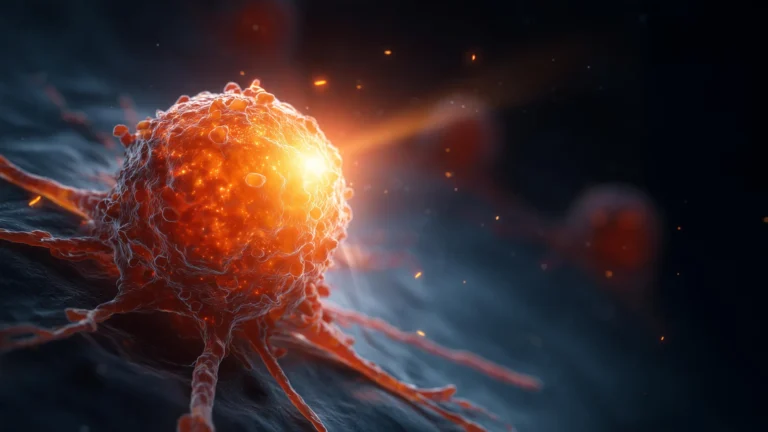
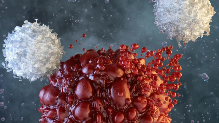
In the silent, relentless war against infection and malignancy, cytotoxic T lymphocytes (CTLs) act as the elite shock troops of the human immune system. These specialized "killer" cells possess the lethal ability to identify and eradicate rogue elements—whether they be virus-infected cells or malignant tumors—with surgical precision. Central to this process is the "immune synapse," a highly orchestrated contact zone that serves as the gateway for the delivery of toxic molecules.
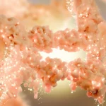
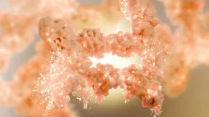
For decades, the mechanics of this interface have been the subject of intense study, yet the nanoscopic choreography occurring within the synapse remained largely obscured by the limitations of traditional imaging. Now, a collaborative breakthrough by researchers at the University of Geneva (UNIGE) and the Lausanne University Hospital (CHUV) has shattered these barriers. By deploying an innovative imaging technique known as cryo-expansion microscopy (cryo-ExM), the team has successfully visualized the three-dimensional, near-native internal architecture of these cells. This discovery, recently published in Cell Reports, not only deepens our understanding of cellular biology but also provides a vital roadmap for the next generation of immuno-oncology therapies.
Chronology of a Breakthrough: From Specimen to Insight
The pursuit of a clearer view of the immune synapse has been a long-standing challenge in cellular biology. Historically, researchers faced an agonizing trade-off: they could achieve high resolution at the cost of distorting the cell’s natural architecture, or they could preserve the cell’s shape while sacrificing the ability to see the fine details of its internal machinery.
The Limitations of Tradition
The primary obstacle was sample preparation. Conventional methods, such as chemical fixation and dehydration, often cause delicate intracellular components to collapse or shift, leading to artifacts that misrepresent the cell’s true state. Furthermore, standard light microscopy often lacked the resolving power to peer into the dense, crowded environment of the synapse.
The Cryo-Expansion Paradigm
To bypass these hurdles, the research team—supported by the ISREC Foundation TANDEM program—integrated two sophisticated technologies: vitrification and physical expansion.
- Instantaneous Freezing: The process begins with rapid cryo-fixation, where cells are frozen in a "vitreous state." By preventing the formation of ice crystals, which would otherwise rupture delicate membranes, the researchers effectively "locked" the cell in its biological reality.
- Expansion Microscopy: Once vitrified, the samples are infused with an absorbent hydrogel. This polymer network physically expands the sample, effectively magnifying the distance between cellular structures. This process allows scientists to use conventional microscopes to visualize nanometer-scale details that were previously invisible.
By combining these methods, the team effectively created a "high-definition" map of the immune synapse, revealing features that had remained hidden under previous investigative paradigms.
Supporting Data: The Architecture of the "Dome"
The study’s findings provide a granular look at the structural mechanics of the CTL-target cell interaction. According to the research, the immune synapse is far from a flat interface; rather, it is a complex, dynamic structure.
The Membrane Dome
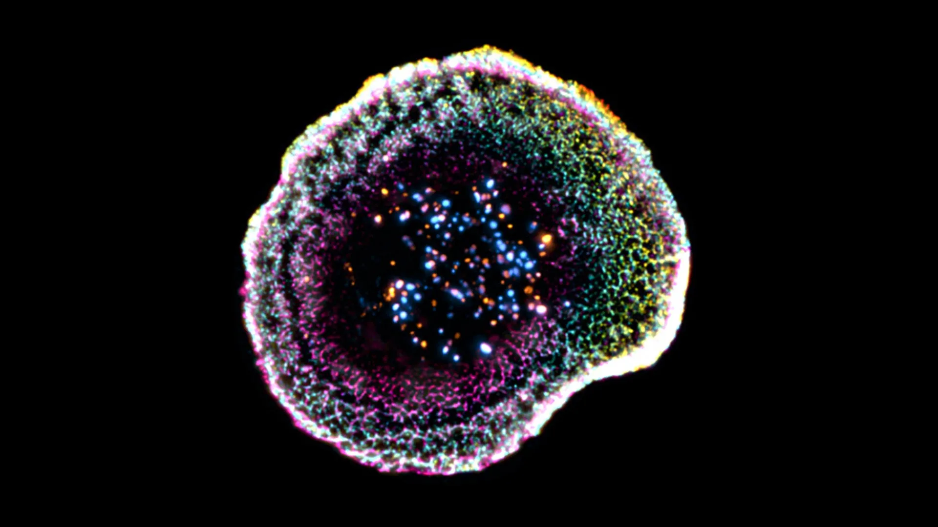
One of the most striking revelations was the discovery of a distinct membrane "dome" formed at the site of contact between the lymphocyte and the target cell. Florent Lemaître, a postdoctoral researcher in the Department of Molecular and Cellular Biology at UNIGE and the study’s first author, notes that this dome is not merely a passive byproduct of contact. "Our work reveals that at the point of contact, the membrane forms a kind of dome, whose structure appears to be linked to adhesion interactions and to the internal organization of the cell," Lemaître explained. This structure suggests that the lymphocyte actively remodels its own cytoskeleton to ensure a sealed environment for the delivery of its toxic payload.
The Granule Complexity
The study also provided unprecedented clarity regarding cytotoxic granules—the "ammunition" storage units within the T cell. Researchers observed that these granules exhibit structural heterogeneity. Some possess single, dense cores, while others contain multiple cores where active molecules are concentrated. This diversity in internal organization may indicate a sophisticated, tiered delivery system that the T cell uses to modulate the intensity of its attack based on the nature of the target.
Official Responses and Expert Perspectives
The project represents a significant triumph of interdisciplinary cooperation between academic researchers and clinical oncologists.
Virginie Hamel, Senior Lecturer at UNIGE’s Department of Molecular and Cellular Biology, emphasized the significance of the methodology. "This technique involves instantaneously freezing cells at very high speed, placing them in a so-called vitreous state… The samples are then physically expanded using an absorbent hydrogel, making it possible to observe their internal organization with great precision while maintaining their near-native architecture."
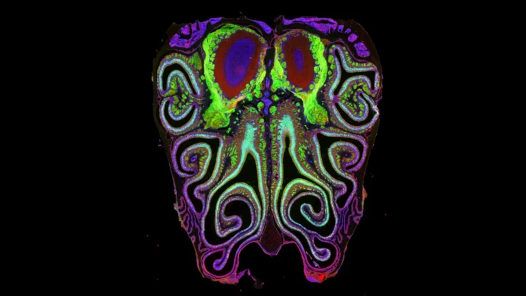
The clinical relevance of these findings was underscored by Dr. Benita Wolf, Chief Resident and Associate Researcher in the Department of Clinical Oncology at CHUV. Dr. Wolf, who co-led the study, highlighted the importance of moving beyond lab-grown cell lines. "We extended this approach to human tumor tissues, making it possible to directly observe T lymphocytes infiltrating tumors and their cytotoxic machinery at the nanometer scale," Dr. Wolf stated. By verifying these mechanisms within actual clinical samples, the team has bridged the gap between basic cellular biology and translational medicine.
Implications for Immuno-Oncology
The implications of this research for the future of cancer treatment are profound. Current immunotherapy strategies, such as CAR-T cell therapy, rely on the ability of engineered T cells to find and kill cancer cells. However, clinical success is often hampered by the "exhaustion" of T cells or their inability to effectively penetrate the hostile tumor microenvironment.
Refining Treatment Strategies
Understanding the structural constraints of the immune synapse allows researchers to ask new questions:
- Why do some T cells fail to form a stable "dome" on tumor cells?
- Can we modify the internal structure of cytotoxic granules to improve the efficacy of engineered T cells?
- How does the physical environment of a solid tumor alter the structural integrity of the immune synapse?
By providing a three-dimensional, nanometer-scale view of these cells in their "near-native" environment, this study offers a new framework for evaluating the effectiveness of immune-based cancer therapies. Researchers can now look for physical "bottlenecks" that prevent successful tumor clearance, potentially leading to the development of drugs or genetic modifications that stabilize the synapse or enhance the delivery of cytotoxic granules.
A New Era of Diagnostic Imaging
Beyond therapy, this imaging methodology offers a powerful diagnostic tool. If researchers can correlate specific structural signatures of T cells with clinical outcomes in patients, they may be able to develop predictive models that determine how well a patient’s immune system is responding to a particular treatment. This move toward personalized, mechanism-based medicine is the cornerstone of modern immuno-oncology.
Conclusion: The Path Forward
The collaboration between UNIGE and CHUV has not only pushed the boundaries of what can be seen under a microscope but has also fundamentally altered our perception of the "killer" cell. The immune synapse is no longer a black box; it is an intricate, mechanical machine, capable of being analyzed, understood, and potentially optimized.
As the scientific community moves forward, the ability to visualize these processes in human tumor tissues will be critical. By aligning the mechanical realities of T cell activity with clinical observations, the research team has set the stage for a new wave of innovations. While there is still much to learn about the signaling pathways that govern these structural changes, the work published in Cell Reports provides the foundation upon which the next decade of life-saving cancer treatments will likely be built. In the fight against cancer, clarity is perhaps our most potent weapon, and through cryo-expansion microscopy, that clarity has finally arrived.