









In the landscape of modern medicine, where diagnostic technology has reached unprecedented levels of precision, some conditions have inexplicably slipped through the cracks of clinical consciousness. Among the most enigmatic is visceroptosis—the downward displacement of internal organs from their normal anatomical positions. While pelvic organ prolapse and rectal prolapse are well-documented phenomena, particularly within the Ehlers-Danlos Syndrome (hEDS) community, the broader concept of abdominal organ displacement has largely been relegated to the annals of medical history.
As patients continue to struggle with chronic, unexplained gastrointestinal distress, a growing number of researchers and clinicians are beginning to ask a critical question: Could the "forgotten" diagnosis of visceroptosis be the missing link in understanding the complex systemic symptoms of connective tissue disorders?
Main Facts: Defining the Displacement
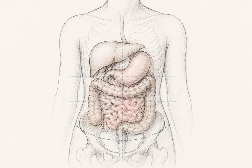
At its core, visceroptosis is a structural diagnosis. The term is derived from "viscera"—the internal organs of the abdominal, thoracic, and pelvic cavities—and "ptosis," which refers to the downward descent of a structure. In a healthy state, abdominal organs are anchored by ligaments, mesenteries, and the supportive tension of the abdominal wall. When these connective tissues lose their integrity—a hallmark of conditions like hEDS—the organs may shift, sagging lower than their intended anatomical coordinates.
However, it is vital to distinguish between anatomic and clinical visceroptosis. Anatomic visceroptosis describes a structural shift that may exist without causing the patient any distress. Clinical visceroptosis, by contrast, occurs when that displacement fundamentally alters the organ’s function, leading to physiological impairment.
The symptoms associated with this condition are as varied as the organs themselves. While pelvic-based ptosis typically presents with pressure, incontinence, or voiding difficulties, abdominal visceroptosis is frequently characterized by:
- Severe, intractable constipation and bloating.
- Early satiety (feeling full after only a few bites of food).
- Chronic abdominal pain.
- Nausea and symptoms mimicking gastroparesis.
These symptoms are often debilitating, yet they frequently fail to show up on standard endoscopic or imaging tests, leaving patients in a cycle of medical dismissal where their physical suffering is incorrectly categorized as psychological.
Chronology: From "Glénard’s Disease" to Obscurity
The history of visceroptosis is a curious tale of medical fashion. In 1885, French physician Dr. Frantz Glénard published seminal work describing patients whose abdominal organs were "dramatically lower" than expected. His observations were so precise that the condition was initially termed "Glénard’s disease."
For the next several decades, visceroptosis was a mainstream medical topic. It was a frequent subject of discourse in premier journals such as The New England Journal of Medicine, JAMA, and The Lancet. Radiologists actively sought signs of displacement, and surgeons developed various "pexy" procedures to reposition organs.
Then, mid-20th century, the discussion abruptly halted. Without a formal consensus on why it fell out of favor, the diagnosis essentially vanished from medical education. Physicians stopped looking for it, and the literature became silent. It was not until the early 21st century, with the rising awareness of connective tissue disorders like Ehlers-Danlos Syndrome, that clinicians began to notice a pattern: patients with hypermobility were frequently reporting the exact symptoms Dr. Glénard described over a century ago.
Supporting Data: The Link to Connective Tissue
The intersection of visceroptosis and hEDS provides the strongest evidence for a resurgence in clinical interest. In patients with hEDS, the collagen—the "glue" of the body—is structurally compromised. This systemic laxity does not stop at the skin or joints; it affects the internal suspensory systems of the body.
Recent research highlights the clinical significance of this link:
- Diagnostic Inclusion: Pelvic organ and rectal prolapse are already recognized as potential complications within the 2017 international diagnostic criteria for hEDS.
- Gastrointestinal Impact: Small-scale case reports have identified instances of gastroptosis (downward stomach displacement) and transverse colon ptosis in hEDS patients.
- Measurable Severity: A significant 2023 study involving 139 patients with chronic, slow-transit constipation revealed that those with higher degrees of transverse colon ptosis were more likely to require surgical intervention. While this study was not exclusive to the EDS population, it serves as a proof-of-concept that organ position is directly correlated to the severity of functional gastrointestinal disease.
Despite these findings, researchers caution against assuming a simple cause-and-effect relationship. Gastrointestinal health in hypermobile patients is multifactorial, involving disorders of gut-brain interaction, autonomic nervous system dysfunction, and pelvic floor weakness. Determining whether a "ptotic" organ is the primary driver of pain or merely an incidental finding remains the primary challenge for modern gastroenterologists.
Official Responses and Clinical Challenges
The current medical establishment remains hesitant to embrace visceroptosis as a standalone diagnosis. The lack of standardized imaging protocols is the most significant hurdle. Current diagnostic pipelines are optimized to detect tumors, ulcers, or inflammatory bowel disease (IBD). They are not designed to assess "anatomical alignment" while the patient is in a standing or functional position.
Because of this, many patients report a familiar, frustrating trajectory: they are diagnosed with "Functional Dyspepsia" or "Irritable Bowel Syndrome (IBS)," told that their tests are normal, and directed toward psychological support. This "neurasthenic" labeling—a term used in the 1880s—mirrors the modern dismissal of patients with "somatic" symptoms.
However, the tide is turning. As advocacy groups like Chronic Pain Partners highlight the disconnect between patient experience and current diagnostic standards, there is a growing push for "functional imaging." This involves using upright MRIs or dynamic fluoroscopy to observe how organs behave under the influence of gravity—a stark departure from the traditional supine (lying down) imaging that currently dominates hospital protocols.
Implications: The Path Toward Treatment
How do we treat a condition that is only just being rediscovered? The current consensus emphasizes a conservative-first approach.
Conservative Management
For many, symptoms can be managed through a multidisciplinary team:
- Physical Therapy: Specialized pelvic floor and core-strengthening physical therapy can help compensate for ligamentous laxity.
- Compression Therapy: The use of specialized abdominal binders or compression garments can provide the external support that internal connective tissues are failing to supply.
- Dietary Modulation: Small, frequent meals are often recommended for those with gastroptosis to reduce the weight load on the stomach.
The Surgical Dilemma
Surgery remains a controversial and high-stakes option. While "pexies" (surgical fixations) were common in the early 20th century, they carry significant risks today, particularly for those with EDS. Connective tissue fragility can lead to poor wound healing, suture failure, and the recurrence of prolapse. Furthermore, surgery in an already sensitized nervous system can sometimes exacerbate chronic pain. Consequently, surgery is generally reserved for cases where conservative measures have failed and where the anatomical displacement is deemed severe and demonstrably linked to the patient’s primary symptoms.
Conclusion: A Call for Clinical Nuance
Visceroptosis is a prime example of why medicine must remain humble. What was once a "forgotten" diagnosis is proving to be a highly relevant piece of the puzzle for a modern patient population living with systemic connective tissue dysfunction.
The path forward requires more than just acknowledging that organs can move; it requires the development of standardized diagnostic criteria, a commitment to functional imaging, and a willingness to listen to the "somatic" experiences of patients who have been told their pain has no physical cause. As we move into the latter half of the 2020s, the revival of this diagnosis may finally provide the clarity and validation that thousands of patients have been seeking for decades.
Author: Amy Weintraub, Research Specialist II, Norris Lab
Editor: Jacqueline Teti, Director of Programs, CPP
Date: July 2026