







Every year, the seasonal influenza virus causes between three and five million cases of severe illness, leading to a staggering death toll of up to 650,000 individuals globally. Beyond its seasonal toll, the virus remains a constant, looming threat of pandemic proportions, echoing the devastation of the 1918 Spanish Flu. Despite decades of study, the precise molecular "handshakes" between the influenza virus and its human host have remained elusive—until now.
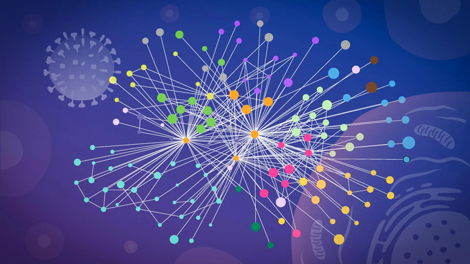
In a landmark study published in Nature Microbiology, researchers from the European Molecular Biology Laboratory (EMBL) in Hamburg, in collaboration with the Leibniz Research Institute for Molecular Pharmacology (FMP) and the Charité – Universitätsmedizin Berlin, have unveiled an unusually detailed map of how influenza A reshapes the interior of infected human cells. By observing protein interactions directly within intact, living cells, the team has bypassed the limitations of traditional laboratory methods, opening a new frontier in the development of next-generation antiviral therapies.
The Molecular Takeover: A Chronology of Infection
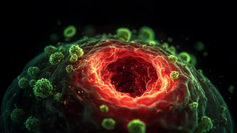
The influenza virus is a master of subversion. Upon entering a human cell, it does not merely occupy space; it systematically dismantles the host’s internal governance. The process begins when the virus sheds its viral RNA, which carries the genetic blueprints for building new viral proteins. These proteins then fan out across the host cell, hijacking its molecular machinery and converting the cell into a high-capacity factory for viral replication.
For years, scientists have understood the "what" of this process—that the cell is repurposed—but the "how" remained obscured. The difficulty lies in the sheer complexity of the intracellular environment. To understand how the virus orchestrates this takeover, researchers must identify which viral proteins dock with human proteins, the precise spatial coordinates of these contacts, and the functional consequences of these unions.
Historically, tracking these interactions in real-time was nearly impossible. Most conventional biochemical techniques required scientists to rupture or "lyse" the cells to analyze their contents. This invasive process often proved destructive, creating an artificial environment where proteins that never interacted in nature suddenly collided, while transient or fragile interactions simply vanished. The resulting data, therefore, was often a blurred snapshot of a distorted reality.
Breaking the Barrier: The Innovation of In-Cell XL-MS
The breakthrough came when the EMBL researchers teamed up with Boris Bogdanow and Fan Liu at the FMP in Berlin. The collaborators had been refining a specialized version of cross-linking mass spectrometry (XL-MS)—a technique that "freezes" protein interactions in place—specifically adapted for infected, living cells.
Overcoming Technical Limitations
Unlike traditional mass spectrometry, this in-cell approach preserves the spatial integrity of the cell. By chemically "cross-linking" proteins that are in close proximity before the cell is broken down for analysis, the researchers could capture snapshots of the virus-host interface exactly as it existed during the active infection.
"XL-MS allows us to capture protein-protein interactions directly in infected intact cells, while also providing structural information about how these interactions are happening," explains Boris Bogdanow, now a Junior Research Group Leader at the Institute of Virology at Charité – Universitätsmedizin Berlin. "This gives us insight into the interface between the virus and the human cell and may, through structural modeling, help identify actionable targets for future pharmaceutical interventions."
Integrating AlphaFold for Structural Precision
The experimental data provided by XL-MS served as the foundational bedrock for the next phase of the research: computational modeling. The team utilized a modified version of AlphaFold, the Nobel Prize-winning protein structure prediction algorithm.
By feeding the experimental cross-linking data directly into the AlphaFold architecture, the researchers could effectively "constrain" the models. "The key advantage of the modified AlphaFold approach is that it allowed us to feed our experimental cross-linking data directly into the structural modeling," notes Jan Kosinski, Group Leader at EMBL Hamburg and the Centre for Structural Systems Biology (CSSB). "This tells the model which parts of the viral and host proteins are close to each other inside infected cells. This was especially useful for virus-host complexes, which are often difficult to predict reliably."
Two Primary Strategies of Viral Hijacking
The study identified two distinct and highly effective strategies that influenza A utilizes to gain total control over the host cell’s resources.
1. Manipulating the Protein Processing Network
The first strategy centers on hemagglutinin, a protein located on the surface of the influenza virus. Hemagglutinin is the virus’s "key," allowing it to attach to and enter host cells. Once inside, the protein must be correctly folded and modified within the cell’s internal transport system—a complex network of compartments known as the secretory pathway.
The research revealed that the virus actively recruits specific human proteins to facilitate the folding and modification of hemagglutinin. By mapping these interactions, the team identified several host proteins whose functions were previously poorly understood, highlighting how the virus exploits host cellular machinery to ensure its own surface proteins are "battle-ready" for the next round of infection.
2. The Dissolution of Paraspeckles
The most startling discovery involved "paraspeckles," which are small, droplet-like compartments residing within the cell nucleus. The team observed that in every cell line and every influenza strain tested, infection caused these structures to dissolve.
"What surprised us most was the paraspeckles," says Iuliia Kotova, the study’s first author and a former predoctoral fellow at the Kosinski Group. "Watching these tiny organelles in the nucleus dissolve, consistently across every cell line and every flu strain we tested, told us this isn’t a side effect of infection—it might be a strategy."
The dissolution of paraspeckles appears to be a dual-purpose maneuver. First, it releases RNA-binding proteins that were previously sequestered inside the droplets, which the virus likely repurposes to boost its own replication. Second, it may be a tactical strike against the host’s immune response. Paraspeckles are known to play a role in cellular stress responses and the regulation of antiviral genes; by dismantling them, the virus effectively blinds the cell’s internal alarm system, preventing it from mounting a coordinated defense.
Implications for Future Medicine
The implications of this "mapping in context" approach are vast. By identifying exactly which proteins are being hijacked and how, researchers can move toward developing precision medicines—drugs that block these specific interactions without causing the broad, toxic side effects often associated with current antiviral therapies.
A Blueprint for Pandemic Preparedness
While the current study focused on a laboratory-adapted strain of influenza, the methodology is designed to be highly scalable. The team believes this approach can be applied to investigate more dangerous viruses, such as H5N1 (avian influenza), which poses a significant pandemic threat.
"Although this study has focused on a lab-adapted strain, this work lays the groundwork to apply the methodology to viruses of potential pandemic relevance," says Bogdanow. "It allows us to uncover the interaction networks that support their multiplication in human cells, providing us with a clearer map for future pharmaceutical interventions."
Broadening the Horizon
The success of this collaboration—which spanned three institutions and integrated expertise from proteomics, computational biology, and advanced microscopy—demonstrates the power of interdisciplinary research. As Jan Kosinski concludes, "While the exact host factors and mechanisms often differ from virus to virus, we think our overall approach—combining in-cell cross-linking, structural modeling, and targeted cell-biology follow-up—remains broadly applicable."
As the world continues to grapple with the persistent threat of respiratory viruses, this detailed molecular map provides more than just scientific insight; it provides a strategic manual. By understanding the intricate steps influenza takes to rewrite the rules of the cell, scientists are now better positioned than ever to interrupt the viral cycle, potentially turning the tide in the perpetual battle between human immunity and viral evolution.