










In a medical landscape where chronic stress has long been regarded as an invisible, subjective burden, a team of researchers from the Johns Hopkins University School of Medicine has achieved a breakthrough that promises to change how clinicians diagnose and manage the physical consequences of our modern, high-pressure lives. By leveraging advanced deep-learning artificial intelligence, researchers have successfully identified the first-ever biomarker of chronic stress that can be visualized and quantified through standard medical imaging.
The findings, set to be presented at the upcoming annual meeting of the Radiological Society of North America (RSNA), represent a monumental shift in diagnostic medicine. For the first time, physicians may be able to look at a routine CT scan and objectively measure the long-term physiological toll that stress has taken on a patient’s body, potentially transforming how we approach preventative cardiology and systemic wellness.
The Physiological Burden of the Invisible
Chronic stress is far more than a psychological state; it is a systemic physiological phenomenon. According to the American Psychological Association (APA), prolonged exposure to stressors triggers a cascade of hormonal and inflammatory responses that can degrade physical health over time. From the persistent elevation of blood pressure and the suppression of the immune system to the exacerbation of sleep disorders and muscle tension, the body’s "allostatic load"—the cumulative wear and tear resulting from chronic stress—is a known driver of severe medical conditions, including obesity, major depression, and cardiovascular disease.
Historically, however, quantifying this burden has been a clinical challenge. Practitioners have relied primarily on patient self-reporting through questionnaires or cumbersome laboratory tests, such as repeated salivary cortisol sampling or blood work, to gauge stress levels. These methods often provide only a "snapshot" of a patient’s state at a single moment in time, failing to capture the cumulative damage that occurs over years.
Chronology of the Breakthrough
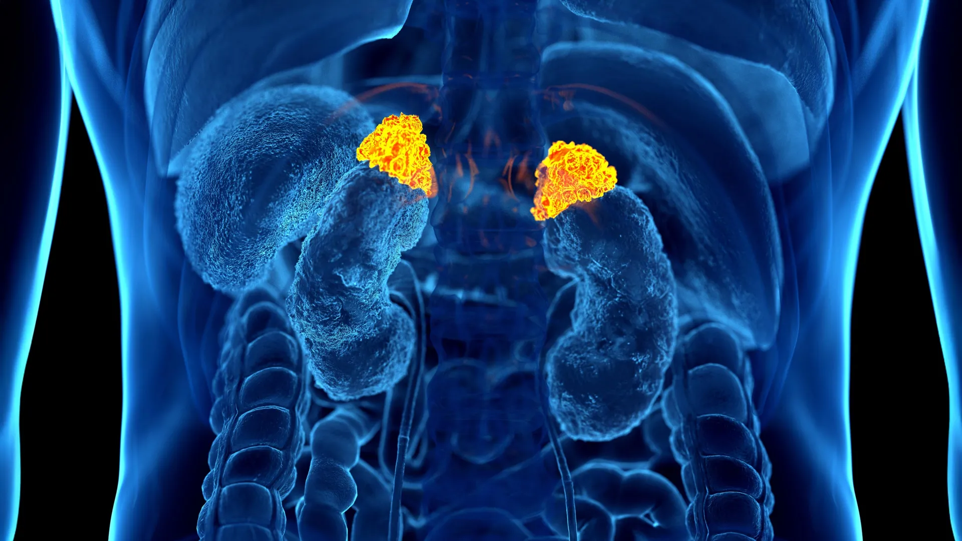
The journey to this discovery began with the recognition of a common, untapped data source: the millions of chest CT scans performed annually in the United States. Dr. Elena Ghotbi, a postdoctoral research fellow at Johns Hopkins, led the initiative to develop a deep-learning model capable of automatically segmenting and measuring the adrenal glands—the organs responsible for the body’s "fight or flight" hormonal response.
- Development Phase: Dr. Ghotbi and her team trained a deep-learning algorithm to identify and calculate the precise volume of the adrenal glands from existing, routine chest CT datasets.
- Cohort Analysis: The model was tested against data from 2,842 participants in the Multi-Ethnic Study of Atherosclerosis (MESA). This cohort was selected specifically because it contained a unique trifecta of data: high-quality chest CT imagery, validated psychosocial stress questionnaires, and comprehensive biochemical markers of allostatic load.
- Validation: The researchers correlated the AI-derived Adrenal Volume Index (AVI) against the participants’ cortisol levels (collected eight times daily over two days) and their physical health metrics, including blood pressure, glucose, and heart rate.
- Outcome Correlation: Finally, the team mapped the AVI results against the participants’ long-term clinical outcomes, tracking occurrences of heart failure and mortality over a follow-up period of up to 10 years.
Supporting Data: Defining the Adrenal Volume Index (AVI)
The study introduced the "Adrenal Volume Index" (AVI), a metric calculated by dividing the adrenal gland volume (cm³) by the square of the patient’s height (m²). This normalization allows for a standardized assessment of adrenal enlargement, which the researchers posited acts as a long-term barometer for chronic stress.
The data revealed a striking consistency between the AI’s measurements and the patients’ lived experiences. Participants who reported higher levels of perceived stress on standardized questionnaires demonstrated consistently higher AVI values. Furthermore, the AI-measured adrenal enlargement showed a statistically significant link to higher total cortisol exposure and elevated peak cortisol levels.
Perhaps most critically, the study linked the AVI to structural heart changes. Elevated AVI was associated with a higher left ventricular mass index, a key indicator of cardiac strain. The researchers found that for every 1 cm³/m² increase in the Adrenal Volume Index, there was a measurable increase in the risk of heart failure and cardiovascular-related death. This makes the AVI not just a marker of stress, but a predictive tool for life-threatening clinical outcomes.
Official Responses and Expert Perspective
The significance of these findings lies in the practicality of the technology. By utilizing imaging that is already being performed for other clinical reasons—such as lung cancer screenings or thoracic diagnostic assessments—the medical community gains a "free" window into the patient’s stress history.
"For the first time, we can ‘see’ the long-term burden of stress inside the body," said Dr. Shadpour Demehri, a professor of radiology at Johns Hopkins and the senior author of the study. "Until now, we haven’t had a way to measure and quantify the cumulative effects of chronic stress, other than questionnaires or surrogate serum markers. This approach leverages widely available imaging data and opens the door to large-scale evaluations of the biological impact of chronic stress."
Dr. Teresa E. Seeman, a professor of epidemiology at UCLA and a co-author of the study, emphasized the longitudinal importance of the work. "For over three decades, we’ve known that chronic stress can wear down the body across multiple systems," Dr. Seeman noted. "What makes this work so exciting is that it links a routinely obtained imaging feature—adrenal volume—with validated biological and psychological measures. It’s a true step forward in operationalizing the cumulative impact of stress on health."
Clinical Implications: The Future of Preventative Care
The integration of this AI-driven biomarker into standard radiology workflows holds profound implications for clinical practice.
Enhanced Risk Stratification
Cardiologists could potentially use the AVI as an additional layer of data when evaluating a patient’s cardiovascular risk profile. If a scan reveals an enlarged adrenal gland, it could signal that the patient is experiencing a level of chronic physiological stress that standard blood tests might miss, prompting earlier interventions or more aggressive lifestyle modifications.
Shifting from Reactive to Proactive
Currently, medical intervention for stress-related illnesses often begins only after the development of symptoms or a major cardiac event. The ability to identify high-risk, "high-stress" individuals before they become symptomatic allows for a proactive approach. Clinicians could recommend stress-reduction programs, nutritional counseling, or mental health support based on the objective evidence provided by the scan.
Reducing Healthcare Costs and Radiation
Because this marker is derived from existing CT scans, it requires no additional radiation exposure, no extra laboratory tests, and no additional burden on the patient. It maximizes the utility of medical data that is already being paid for and performed, making it a highly cost-effective tool for large-scale public health screenings.
Conclusion: A New Standard for Wellness
The research presented by the Johns Hopkins team marks the transition of chronic stress from an abstract, often overlooked clinical variable into a tangible, measurable medical entity. As the Radiological Society of North America prepares to host the official presentation of these findings, the broader medical community is looking toward a future where "stress" is no longer just something a patient mentions in passing during a check-up, but a quantifiable metric that can guide the course of a patient’s long-term cardiovascular health.
By bridging the gap between imaging, endocrinology, and cardiology, Dr. Ghotbi and her colleagues have provided the field with a powerful, data-driven instrument. The Adrenal Volume Index may well become a standard component of future diagnostic reports, ensuring that the invisible toll of our fast-paced world is no longer ignored by the eyes of modern medicine.