








Congenital heart disease (CHD) remains one of the most pressing challenges in modern neonatology. Affecting roughly two out of every 100 newborns worldwide, these structural abnormalities represent a staggering global health burden, with millions of children born annually requiring complex, life-altering medical interventions. Despite decades of intense study, the precise molecular triggers that cause the human heart to deviate from its healthy developmental path during gestation have remained elusive.
However, a breakthrough study from the University of Copenhagen, recently published in the scientific journal PLOS Biology, has identified a previously unknown cellular mechanism that acts as a foundational "cog" in the complex machinery of organ formation. By uncovering how a microscopic cellular antenna guides heart development, researchers have provided a new framework for understanding not just heart defects, but a wide array of syndromic genetic disorders.
The Primary Cilium: A Microscopic Command Center
At the heart of this discovery is the "primary cilium"—a tiny, hair-like projection found on the surface of nearly every cell in the human body. For years, these structures were dismissed by some as vestigial, but they are now recognized as sophisticated sensory organelles.
"Primary cilia act as microscopic antennas," explains Professor Søren Tvorup Christensen of the Department of Biology at the University of Copenhagen. "They extend from the cell surface to sense and interpret chemical signals from the surrounding environment. These signals are the ‘go’ or ‘stop’ commands that dictate whether a stem cell should divide, move, differentiate into a specific tissue, or even undergo programmed cell death."
During the critical window of embryonic development, these antennas are tasked with coordinating the formation of vital organs, including the heart, brain, and skeleton. The research team discovered that three specific proteins—TAK1, TAB2, and PKA-Cα—operate as a specialized signaling hub within these cilia. This hub acts as a gatekeeper, processing the molecular instructions that transform generic stem cells into specialized heart muscle cells. When this communication is disrupted by genetic mutations, the "antenna" fails, leading to the structural malformations characteristic of CHD.
A Multi-Year Investigation: From Genetic Data to Biological Reality
The discovery was the result of a rigorous, multi-disciplinary approach that spanned several years. The team’s methodology was designed to bridge the gap between abstract genetic data and observable biological phenomena.
Phase 1: Identifying the Genetic Signature
The researchers began by analyzing genetic data from several thousand patients living with congenital heart defects. By comparing the genetic profiles of these patients against healthy control groups, the team identified rare mutations that appeared with significantly higher frequency in the patient population. These variants were flagged as high-probability contributors to the underlying disease.
Phase 2: Functional Validation in Zebrafish
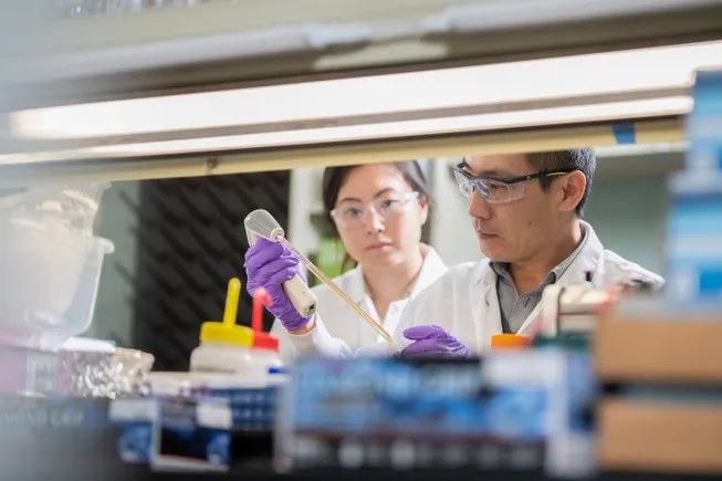
To determine the biological impact of these mutations, the team utilized genetic engineering to recreate the identified human mutations in zebrafish. Zebrafish are an ideal model for heart development, as their embryonic heart formation mirrors many aspects of human organogenesis. The results were clear: the engineered mutations consistently interfered with heart function and architecture, confirming that these genes were not just associated with the disease—they were driving it.
Phase 3: Cellular Modeling
Finally, the researchers utilized human and mouse stem cells in laboratory cultures. By studying these cells in a controlled environment, they were able to observe the signaling hub in real-time, documenting exactly how the communication pathways collapsed when the TAK1, TAB2, or PKA-Cα proteins were disrupted.
"We investigated the mechanism from many different angles and using many different methods," says Professor Lars Allan Larsen of the Department of Cellular and Molecular Medicine. "All our findings converged on the same conclusion: the primary cilium is a critical component of the heart’s developmental blueprint. We are reasonably confident this mechanism functions identically in humans."
Supporting Data: The Global Burden of CHD
The scale of the challenge addressed by this study cannot be overstated. According to the World Heart Federation and the Danish Heart Foundation, the prevalence of congenital heart disease is a global public health crisis:
- Annual Incidence: Approximately 2.3 to 2.5 million newborns are affected by CHD each year worldwide.
- Living Population: As of 2023, an estimated 16 million people are living with the long-term consequences of congenital heart defects.
- Syndromic vs. Non-Syndromic: The study specifically focused on "syndromic" congenital heart disease, where heart defects occur alongside abnormalities in other organ systems. In non-syndromic cases, the heart is the sole focus of the defect.
The researchers believe that because the primary cilium is a universal cellular structure, the failure of this signaling mechanism likely accounts for the multisystem complications seen in syndromic patients, potentially linking heart defects to issues in the kidneys, brain, and skeletal development.
Official Perspectives: Implications for Future Medicine
The research team, led by Professors Larsen and Christensen, emphasizes that this discovery is not merely an academic milestone; it is a clinical roadmap.
"This finding changes our understanding of why some congenital heart defects arise," says Professor Larsen. "By identifying the specific proteins involved in this ciliary signaling hub, we have identified a target for future diagnostic and therapeutic interventions."
The implications extend far beyond the heart. Because many rare genetic disorders—known collectively as "ciliopathies"—are caused by defects in the primary cilium, this research could unlock secrets behind diseases that have long remained medical mysteries.
"When the ciliary mechanism fails, it typically affects the development of several other organs as well," notes Professor Christensen. "This may explain why some patients with congenital heart disease also have related conditions affecting the brain or kidneys. This mechanism provides a unifying explanation for a range of diseases that we have previously struggled to categorize."
Looking Ahead: The Path to Early Diagnosis and Targeted Therapy
While the study offers a powerful explanation for the origins of CHD, the researchers remain cautious about the timeline for clinical application. Because the evidence is built upon genetic associations and laboratory models, the team acknowledges that further research is required to map the exact, patient-specific pathways of these mutations in human clinical settings.
However, the mood in the scientific community is optimistic. By moving the focus from broad genetic markers to specific, localized signaling hubs within the cell, the team has significantly narrowed the search field for clinicians.
"Many rare genetic diseases are caused by changes in genes that affect ciliary function, yet the underlying mechanisms have remained poorly understood," Professor Larsen concludes. "This new knowledge may eventually make it easier to identify patients at high risk very early in their development and, in the future, develop targeted treatments that could mitigate the severity of these conditions."
The study, which involved a vast team of researchers from the University of Copenhagen, represents a significant leap forward in the field of developmental biology. As scientists continue to "unmask" the hidden functions of our cells, the mystery of the heart’s formation is becoming, one cog at a time, much clearer.
Research Contributors
The study was a massive collaborative effort involving experts from the University of Copenhagen, including:
Søren Tvorup Christensen, Lars Allan Larsen, Canan Doganli, Oskar Kaaber Thomsen, Daniel A. Baird, Yeasmeen Ali, Menachem V. K. Sarusie, Line Jeanett Jessen, Pauline Munck Truelsen, Johanne Bay Mogensen, Maria Schröder Holm, Lorenzo Buttò, Maria Diamanti, Jindřiška Leischner Fialová, and Lotte Bang Pedersen.
The full research article, titled "The Primary Cilium as a Signaling Hub in Heart Development," is available in the latest issue of PLOS Biology.