









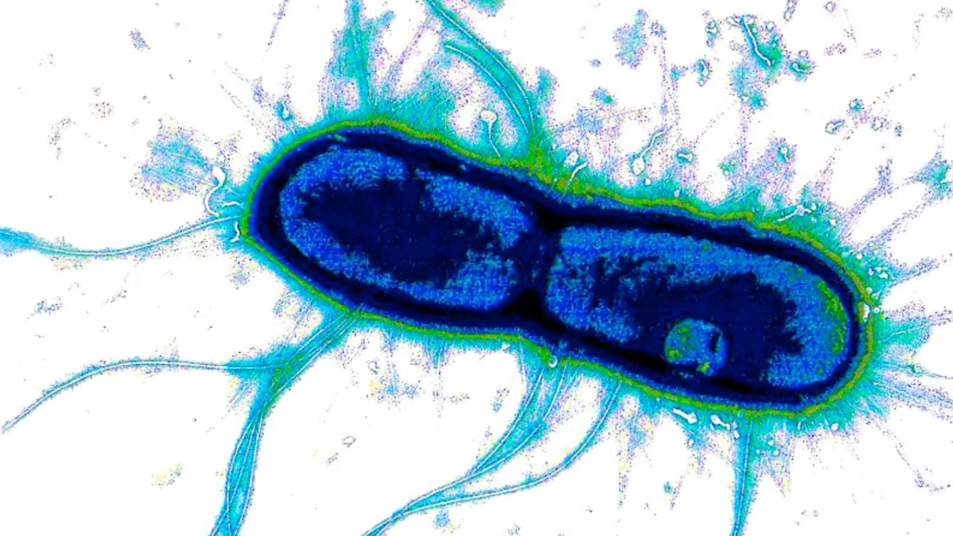
For decades, the global medical community has grappled with an invisible, persistent, and lethal adversary: severe diarrheal disease. Caused primarily by Enterotoxigenic E. coli (ETEC) and Shigella, these pathogens are responsible for hundreds of millions of infections annually. They remain a leading cause of mortality in children under five, particularly in low- and middle-income nations, and represent a significant health burden for travelers and military personnel globally.
Despite the staggering human toll, the quest for a vaccine has been repeatedly thwarted by the evolutionary cunning of these bacteria. Their surface proteins—the traditional targets for vaccine development—are highly polymorphic, meaning they vary wildly between strains. A vaccine that works against one variant may be rendered useless against the next.
However, a groundbreaking study published June 15 in the Proceedings of the National Academy of Sciences (PNAS) has unveiled a fundamental biological vulnerability shared by these disparate pathogens. Researchers at Washington University School of Medicine in St. Louis, in collaboration with the University of Missouri and the International Centre for Diarrhoeal Disease Research, Bangladesh (icddr,b), have identified a common "Achilles’ heel." By targeting the enzymatic machinery these bacteria use to breach the human gut, scientists may finally have a pathway to a single, cross-protective vaccine.
The Mechanics of Invasion: How Pathogens Breach the Gut
To understand the significance of this discovery, one must first understand the battlefield. The human intestine is lined with a thick, viscous layer of mucus—a sophisticated, multi-functional barrier. This mucus acts as a biological filter, separating the body’s delicate intestinal tissues from the trillions of microbes residing in the gut lumen. It serves as a sanctuary for beneficial bacteria while simultaneously keeping invasive pathogens at bay.
Pathogens like ETEC and Shigella have evolved a specialized "drill" to bypass this defense. They secrete specific enzymes that act as molecular shears, cutting through the glycoproteins that provide the mucus layer with its structural integrity. Once this barrier is compromised, the bacteria gain direct access to the epithelial cells lining the intestine, where they release toxins that trigger the catastrophic fluid loss characteristic of severe diarrhea.
For years, Dr. James M. Fleckenstein, a professor of medicine in the Division of Infectious Diseases at WashU Medicine and co-senior author of the study, has focused on this early stage of infection. His laboratory previously identified an enzyme in ETEC called EatA, which specifically degrades the protein components of intestinal mucus.
The new research expands this understanding significantly. The team discovered that Shigella and other related diarrheal pathogens produce two enzymes, SepA and Pic, which are functionally and structurally related to EatA. Because these enzymes are critical for the pathogen’s ability to establish an infection, they represent a "bottleneck" in the bacterial life cycle. By disabling these enzymes, the host’s immune system can effectively keep the bacteria trapped within the harmless mucus layer, where they can be flushed out of the system without ever causing illness.
A Chronology of Discovery: From Clinical Observations to Structural Biology
The path to this discovery was not linear; it was a multi-year effort that bridged clinical observation in Bangladesh with high-resolution structural biology in Missouri.
Phase 1: Clinical Insights (2010s – 2022)
The foundation of the research began with longitudinal studies of children in Dhaka, Bangladesh. The team observed a striking correlation: children who developed natural antibodies against the EatA enzyme were significantly less likely to suffer from severe, symptomatic ETEC infections. Conversely, those lacking these specific antibodies were at a much higher risk of disease. This clinical evidence provided the "proof of concept" that targeting these enzymes could confer immunity.
Phase 2: Antibody Isolation (2022 – 2023)
Building on this, Dr. Fleckenstein partnered with Dr. Ali Ellebedy, a renowned immunologist at WashU Medicine. Using blood samples from individuals who had been naturally exposed to ETEC or who had volunteered for controlled exposure studies, the team isolated specific antibodies. They found that these antibodies, originally identified for their interaction with EatA, possessed a remarkable property: they could also neutralize the SepA and Pic enzymes found in Shigella.
Phase 3: Structural Mapping (2023 – 2024)
To confirm exactly how these antibodies worked, the team utilized cryo-electron microscopy (cryo-EM). Led by Dr. David P. Buckley and Dr. Zachary Berndsen at the University of Missouri, the team flash-froze the enzyme-antibody complexes to capture them in atomic-level detail. The images revealed that the antibodies were binding to a highly conserved region—a shared "signature"—present across all three enzymes (EatA, SepA, and Pic). This explained why a single antibody could neutralize multiple pathogens: the bacteria had shared a common tool, and that tool contained a structural vulnerability that the immune system could exploit.
Supporting Data: The Power of Neutralization
The data generated by the team provides a compelling argument for the feasibility of a combination vaccine. By targeting a conserved enzymatic region, the researchers have effectively bypassed the "variability problem" that has stalled previous vaccine efforts.
The study indicates that these enzymes are not merely accessories to infection; they are essential virulence factors. In the absence of functional EatA/SepA/Pic activity, the bacteria are essentially "blunted," unable to penetrate the mucus barrier to reach the sensitive intestinal wall. Because the antibodies target the functional enzymatic site rather than the surface proteins that mutate rapidly, the evolutionary pressure on the bacteria to "escape" these antibodies is significantly higher, potentially leading to more durable immunity.
Furthermore, the researchers emphasize that this strategy is highly targeted. By stopping the bacteria at the mucosal surface, the vaccine would prevent the initiation of the infection cycle without disrupting the broader, healthy microbiome of the gut. This is a significant improvement over broad-spectrum antibiotics, which often wipe out beneficial commensal bacteria, leading to secondary issues like dysbiosis or C. difficile infections.
Official Responses and Expert Perspectives
The research has been met with optimism within the infectious disease community.
"This study establishes EatA as a viable vaccine candidate capable of providing protection across multiple pathogens," said Dr. Zachary Berndsen. "By identifying the key regions of EatA that are targeted by neutralizing antibodies… we’ve established a foundation for rational vaccine design."
Dr. James M. Fleckenstein, reflecting on the broader impact of the work, noted: "For something so common and so deadly to young children, it’s striking that we still don’t have a vaccine for either of these pathogens. What’s exciting here is that we’ve found a kind of Achilles’ heel they share that we might be able to target to protect against both."
The implications are not limited to the developing world. In the United States, ETEC is a frequent cause of foodborne illness and is notoriously difficult to diagnose in clinical settings, as it is often indistinguishable from benign E. coli strains. By creating a vaccine that prevents the mechanism of infection, the medical community could move away from the current, heavy reliance on antibiotics, which is a primary driver of global antimicrobial resistance (AMR).
Future Implications: The Path Toward Clinical Trials
The team is now transitioning from the laboratory bench to the initial stages of vaccine development. The goal is to design a therapeutic that presents this conserved enzymatic region to the immune system, stimulating a robust antibody response that stands ready to intercept pathogens the moment they enter the gut.
While the path to a regulatory-approved vaccine remains long—requiring rigorous safety testing, animal models, and clinical trials—the discovery of this shared vulnerability marks a pivotal shift in strategy. Instead of chasing the constantly shifting "masks" worn by bacterial surfaces, scientists have identified the "tools" the bacteria use to survive.
The potential to save countless lives—particularly among the world’s most vulnerable children—is the driving force behind this work. As the researchers move forward, their findings serve as a powerful reminder of how basic scientific inquiry, combined with advanced structural imaging, can dismantle even the most persistent of public health threats.
This work was supported by the National Institute of Allergy and Infectious Diseases (NIAID) of the National Institutes of Health (NIH) and the Department of Veterans Affairs. It represents a significant step forward in the international effort to curb the burden of diarrheal diseases and demonstrates the immense potential of targeting conserved virulence mechanisms in the fight against antibiotic-resistant bacteria.