










Featured Buzz — January 11, 2026
The landscape of respiratory medicine is undergoing a profound transformation. As technology bridges the gap between diagnostic potential and clinical application, researchers are unveiling new tools that promise to catch chronic diseases earlier, improve outcomes for the most vulnerable infants, and curb the global crisis of antibiotic overuse. From the halls of Mount Sinai to the intensive care units of the University of California, the latest breakthroughs suggest that the future of pulmonary health lies in the convergence of artificial intelligence (AI), biomarkers, and high-resolution imaging.
I. Early Detection: AI-Enhanced ECGs and the COPD Challenge
Chronic Obstructive Pulmonary Disease (COPD) remains one of the world’s leading causes of mortality and morbidity. Frequently underdiagnosed until the disease reaches an advanced stage, COPD often goes unnoticed during routine clinical interactions. However, a groundbreaking study from the Mount Sinai Health System suggests that the solution to this diagnostic delay may be sitting in every physician’s office: the standard electrocardiogram (ECG).
The Methodology of Predictive Analysis
The research team embarked on a massive data-mining project, harnessing the power of deep learning to extract patterns invisible to the human eye. By analyzing 208,231 ECGs from 18,225 patients with confirmed COPD, and comparing them against a control group of 552,771 ECGs from 59,356 age-, sex-, and race-matched individuals, the team trained a Convolutional Neural Network (CNN).
This model was specifically designed to identify subtle electrical variations in cardiac rhythms and wave patterns that correlate with the physiological stressors COPD imposes on the heart and lungs. By utilizing ICD codes as the gold standard for clinical diagnosis, the model was put through rigorous testing across internal and external cohorts, including a dedicated validation cohort of 258 COPD cases.
Implications for Clinical Practice
The researchers are quick to clarify that this technology is not intended to replace spirometry—the current gold standard for COPD diagnosis. Instead, they frame AI-interpreted ECGs as a "pragmatic screening tool."
"Earlier recognition may facilitate timely smoking cessation, targeted therapies, and pulmonary rehabilitation," the authors noted in their publication in eBioMedicine. By integrating this screening into routine cardiac check-ups, healthcare providers could potentially slow disease progression and significantly reduce the long-term healthcare burden associated with late-stage pulmonary failure.
II. Protecting the Most Vulnerable: Lung Ultrasound in NICUs
In the high-stakes environment of the Neonatal Intensive Care Unit (NICU), the decision to remove a very low birth weight (VLBW) infant from mechanical ventilation is a critical juncture. Extubation failure—the need to re-intubate within seven days—is associated with increased mortality and longer hospital stays. Researchers at the University of Chicago have now validated a tool that could make this transition significantly safer: the neonatal-adapted lung ultrasound (LUS).
Chronology of the Clinical Study
The study followed 45 VLBW infants who had been intubated for respiratory distress syndrome. Across these infants, 53 extubation attempts were monitored. The protocol required a standardized lung ultrasound to be performed three to six hours prior to each attempt.
The findings, published in the Journal of Perinatology, indicate that LUS scores are highly predictive of success. Infants who achieved specific ultrasound milestones demonstrated a higher likelihood of breathing independently post-extubation. Furthermore, the study addressed a common clinical question regarding dexamethasone—a steroid often used to facilitate extubation—finding that its use did not artificially inflate LUS scores, thereby validating the scan’s reliability regardless of pharmaceutical intervention.
The Shift Toward Non-Invasive Monitoring
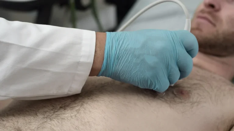
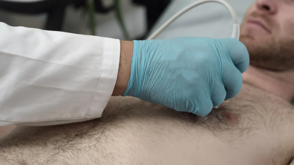
This approach represents a major step toward "radiation-free" bedside diagnostics. Unlike chest X-rays, which expose neonates to ionizing radiation and often require transport out of the incubator, LUS can be performed instantly at the bedside. For clinicians, this means real-time data, less stress on the infant, and more precise decision-making in the delicate hours leading up to life-sustaining milestones.
III. Precision Medicine: Combating Antibiotic Overuse in Critical Care
The misuse of antibiotics is a global health threat, fueling the rise of multidrug-resistant "superbugs." In the ICU, where distinguishing between bacterial pneumonia and other inflammatory conditions is notoriously difficult, clinicians often default to prescribing broad-spectrum antibiotics "just in case." A new diagnostic model developed at the University of California, San Francisco (UCSF), aims to change this paradigm.
The Science of the "Smart" Diagnosis
The UCSF team developed an AI-driven model that combines biological data with clinical context. The biological anchor of this model is FABP4, a gene expressed in lung fluid that plays a key role in modulating inflammation. Crucially, the expression of FABP4 is significantly lower in the presence of a lower respiratory tract infection (LRTI) than in healthy tissue.
By pairing this biomarker with a generative AI analysis of electronic medical records—including chest X-ray reports and physician notes—the model creates a comprehensive picture of the patient’s infection status.
Performance and Data Accuracy
In an observational study involving critically ill adults, the AI model achieved an impressive 96% accuracy rate in diagnosing LRTIs, consistently outperforming experienced ICU clinicians. The potential impact is staggering: researchers estimate that utilizing this tool could reduce the use of inappropriate antibiotics by more than 80%.
"This study suggests that integrating a host biomarker with large language model analysis can improve LRTI diagnosis in critically ill adults," the authors wrote in Nature Communications. The team is currently moving to validate the model for widespread clinical use, with plans to expand the methodology into the diagnosis of sepsis—a leading cause of death worldwide.
IV. Synthesis and Future Outlook
The common thread connecting these three studies is the shift toward "augmented intelligence." We are moving away from an era where clinicians must rely solely on clinical intuition or outdated diagnostic markers. Instead, we are entering a phase where the "hidden" data in our biology (FABP4), our electrical systems (ECG), and our bedside imaging (LUS) can be synthesized by machines to provide a more accurate, personalized, and efficient standard of care.
The Human-AI Partnership
While these technologies offer remarkable promise, they also highlight the necessity of the human element. Whether it is the ICU physician verifying an AI’s diagnosis or the neonatologist interpreting an LUS score, the technology serves as a decision-support system, not a replacement for medical judgment.
Challenges and Ethical Considerations
As these tools move from research papers to clinical deployment, several hurdles remain:
- Integration: Can these AI models be seamlessly integrated into existing Electronic Health Record (EHR) systems without increasing the "alert fatigue" already plaguing clinicians?
- Standardization: As noted with the Mount Sinai ECG study, models trained on specific datasets must be validated across diverse populations to ensure that AI does not perpetuate existing healthcare disparities.
- Regulation: The move toward HIPAA-compliant GPT-4 interfaces for diagnostics marks a new frontier in medical regulation. Ensuring these systems are secure, transparent, and explainable is paramount to maintaining patient trust.
Concluding Thoughts
The advancements reported this January 2026 highlight a vibrant, data-driven future for respiratory medicine. By identifying COPD earlier, making safer decisions for pre-term infants, and utilizing precision diagnostics for pneumonia, the medical community is finding ways to do more with the data we already have. As these models evolve, the focus will likely shift from merely "identifying" disease to "predicting" it—moving medicine from a reactive discipline to one that is fundamentally proactive.
For the patients waiting for answers, these innovations represent more than just academic success; they represent a future where respiratory care is safer, faster, and more effective than ever before.
For further reading on the methodologies and clinical trials discussed above, please refer to the following peer-reviewed publications:
- ECG-based COPD Screening: eBioMedicine
- LUS and Extubation: Journal of Perinatology
- AI-driven LRTI Diagnosis: Nature Communications