











Influenza A remains one of humanity’s most persistent viral adversaries. Responsible for 3 to 5 million cases of severe illness annually and claiming up to 650,000 lives, the virus is a master of biological subversion. It is also the architect of some of history’s most devastating pandemics, most notably the 1918 Spanish Flu. For decades, the primary challenge for virologists has been understanding exactly how the virus—a relatively simple package of RNA and proteins—manages to commandeer the complex machinery of a human cell.
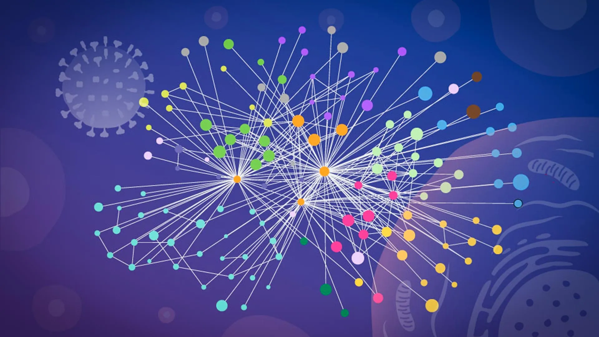
Now, a collaborative team from EMBL Hamburg, the Leibniz Research Institute for Molecular Pharmacology (FMP), and Charité – Universitätsmedizin Berlin has achieved a scientific breakthrough. By utilizing a cutting-edge workflow that captures protein interactions in their native, intact state, researchers have produced an unusually detailed map of the influenza A infection cycle. This study, published in Nature Microbiology, offers not just a static snapshot, but a dynamic view of how the virus rewires host cellular processes to ensure its own replication.
The Problem of Perspective: Breaking the Cell, Losing the Truth
To understand the magnitude of this achievement, one must first understand the limitations of previous methodologies. Historically, "mapping" the interaction between viral proteins and host proteins required a destructive process: researchers would break open infected cells to isolate their contents.
This methodology, while foundational, came with a significant caveat: it fundamentally altered the biological environment. Once the cellular architecture is obliterated, proteins that were once sequestered in different compartments are suddenly allowed to interact, creating "false positives" in the laboratory. Conversely, transient, weak, or highly localized interactions—often the most critical ones for viral replication—are frequently lost in the shuffle. Scientists were essentially trying to understand the blueprint of a building by looking at a pile of rubble.
"The current results are a snapshot of a moment during infection, and it opens the door to studying flu-host interactions across the entire infection cycle," says Jan Kosinski, Group Leader at EMBL Hamburg and the Centre for Structural Systems Biology (CSSB).
A Chronology of Discovery: From Specialized Chemistry to Computational Modeling
The breakthrough came when Kosinski’s team aligned with the expertise of Boris Bogdanow and Fan Liu at FMP Berlin. They had developed a specialized, high-precision version of cross-linking mass spectrometry (XL-MS) designed specifically for the chaotic environment of an infected cell.
The Methodology
- In-Cell Cross-Linking: The team applied chemical cross-linkers directly to living, infected cells. These molecules act like microscopic "staples," locking protein-protein interactions in place at the exact moment of contact.
- Mass Spectrometry Analysis: By stabilizing these interactions, the researchers could then break the cells open and analyze the contents without fear of the protein "partners" drifting apart or forming false associations.
- Structural Integration: To make sense of the vast datasets generated by XL-MS, the team utilized a modified version of AlphaFold, the Nobel Prize-winning protein structure prediction algorithm.
By feeding their experimental cross-linking data directly into the AlphaFold model, the team created a "guided" structural prediction. This informed the algorithm which parts of the viral and human proteins were in close proximity, allowing for a high-fidelity rendering of the virus-host interface. This fusion of experimental data with computational power allowed the team to see the invisible hand of the influenza virus as it gripped its human host targets.
Two Faces of Subversion: How the Virus Takes Control
The findings from this high-resolution mapping revealed two distinct, highly sophisticated strategies employed by the influenza A virus to take control of the host cell.
1. Hijacking the Protein Folding Network
The first discovery centers on hemagglutinin (HA), the protein studding the surface of the flu virus that acts as its "key" to enter human cells. Once inside, the HA protein must be folded, modified, and transported through the cell’s internal assembly line—the secretory pathway.
The researchers tracked HA as it moved through the cell and discovered that the virus actively recruits specific human proteins to act as "chaperones." These host proteins facilitate the folding and modification of HA, ensuring it is ready to be packaged into new, infectious viral particles. Some of these host proteins were previously characterized as having mysterious or poorly defined functions; the study has finally identified their role as essential gear in the influenza replication machine.
2. The Dissolution of Paraspeckles
Perhaps the most surprising discovery involved "paraspeckles"—small, droplet-like structures found within the cell nucleus. The team observed that the influenza A virus causes these structures to dissolve entirely.
"Watching these tiny organelles in the nucleus dissolve, consistently across every cell line and every flu strain we tested, told us this isn’t a side effect of infection—it might be a strategy," says Iuliia Kotova, the study’s first author.
By breaking apart these nuclear compartments, the virus releases a host of RNA-binding proteins that were previously locked away. The virus then appears to co-opt these proteins to bolster its own replication. Furthermore, because paraspeckles are known to play a role in regulating the cell’s stress response and antiviral defenses, their dissolution likely serves a dual purpose: providing the virus with building materials while simultaneously silencing the cell’s internal "alarm system."
Official Perspectives and Collaborative Synergy
The success of this study was fundamentally a triumph of institutional cooperation. The sheer complexity of the data required a cross-disciplinary approach:
- FMP Berlin provided the specialized XL-MS techniques.
- The EMBL Proteomics Core Facility conducted the glycoproteomics analysis.
- The EMBL Compute Cluster handled the intensive AlphaFold modeling.
- CSSB’s Advanced Light and Fluorescence Microscopy (ALFM) Facility provided the high-resolution imaging that confirmed the destruction of the paraspeckles.
Boris Bogdanow, now a Junior Research Group Leader at the Institute of Virology at Charité, underscores the pharmaceutical potential of these findings: "XL-MS allows us to capture protein-protein interactions directly in infected intact cells, while also providing structural information… This gives us insight into the interface between the virus and the human cell and may, through structural modeling, help identify actionable targets for future pharmaceutical interventions."
Implications for Global Health and Pandemic Preparedness
The implications of this research extend far beyond the laboratory strain of influenza used in the study. By successfully mapping the "native context" of virus-host interactions, the team has established a new gold standard for studying viral pathogenesis.
A New Framework for Virology
While every virus has its own unique way of hijacking cellular machinery, the "mapping in context" approach—combining in-cell cross-linking, structural modeling, and targeted cell biology—is a versatile toolkit. The researchers suggest that this methodology could be applied to a wide array of pathogens, including viruses with significant pandemic potential, such as the H5N1 avian influenza strain.
Future Therapeutic Avenues
Current influenza treatments, such as neuraminidase inhibitors, target viral proteins directly. However, viruses mutate rapidly, leading to drug resistance. By identifying the specific human proteins that the virus relies on—such as those involved in HA folding or paraspeckle maintenance—scientists may be able to develop "host-directed" therapies. Because these human proteins are less prone to rapid mutation than viral ones, drugs targeting them could be more durable and effective against evolving viral strains.
As the scientific community looks toward the next potential pandemic, the ability to observe the silent, molecular war inside an infected cell in real-time represents a significant leap forward. By moving from a static, broken-cell view to a dynamic, structural map of the infection process, researchers are finally beginning to see the virus not just as a pathogen, but as an engineer of our own biological systems—and in doing so, they are finding the keys to shutting down that engineering.