







For decades, the study of human vascular biology in a laboratory setting was hampered by a fundamental flaw: the medium did not match the message. Scientists attempting to understand how blood moves through the body—and how disease takes root within that flow—often relied on laboratory models that treated blood vessels as simple, straight, uniform tubes. While these models provided foundational data, they were biologically inaccurate. In the complex reality of the human anatomy, vessels are dynamic, bending, branching, narrowing, and widening in a symphony of fluid mechanics that directly dictates the health of the cardiovascular system.
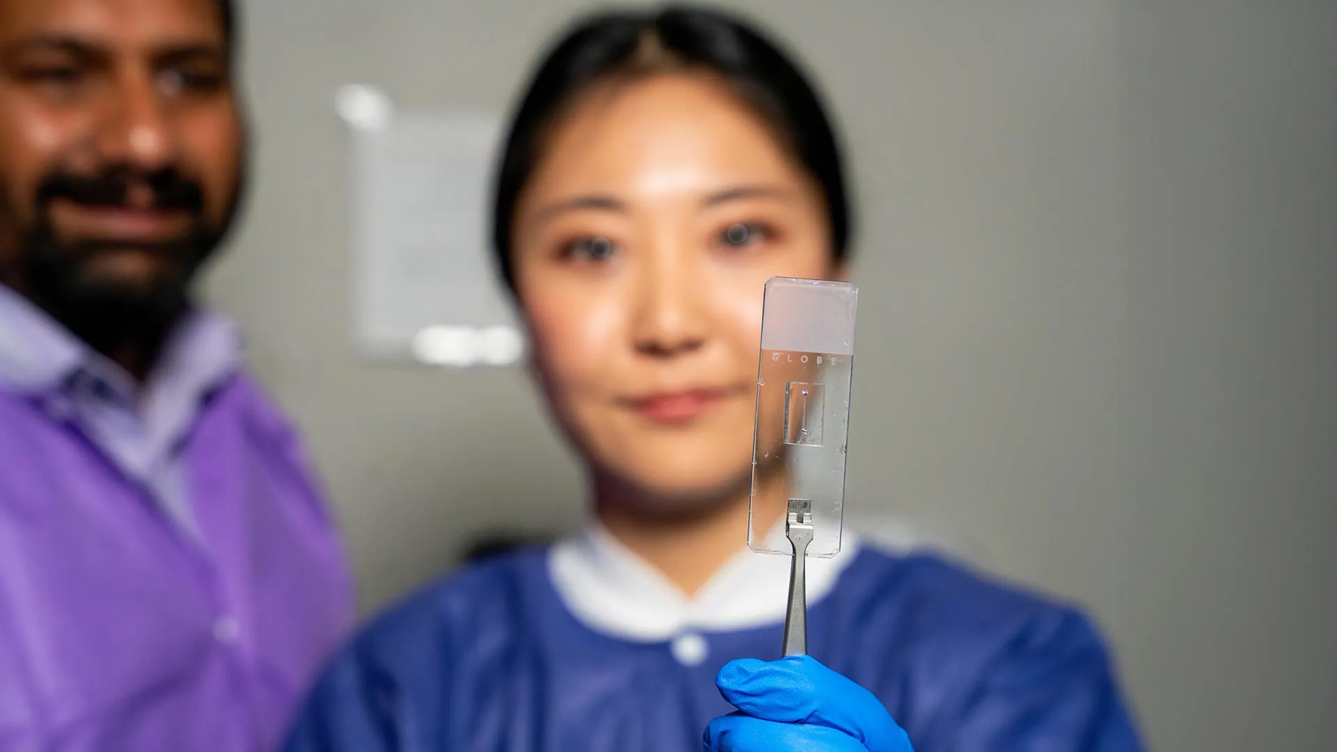
Recognizing this disconnect, researchers in the Department of Biomedical Engineering at Texas A&M University have unveiled a breakthrough in microfluidics. By developing a highly customizable “vessel-chip” system, the team has successfully bridged the gap between simplified lab models and the intricate reality of human physiology. This innovation, spearheaded by master’s student Jennifer Lee under the guidance of Dr. Abhishek Jain, promises to transform how we study vascular disease and test the efficacy of novel pharmaceutical interventions.
The Core Innovation: Moving Beyond Geometry
At the heart of the research, which will be featured on the cover of the May 2025 issue of Lab on a Chip, is the move toward "architectural complexity." Standard microfluidic devices, including an earlier design developed by Dr. Tanmay Mathur in the same lab, were limited by their linear geometry. While useful for basic flow studies, they failed to account for the "shear stress" caused by the irregular shapes of vessels prone to pathology.
Jennifer Lee’s work changes the paradigm. Her vessel-chip system is designed to replicate the specific geometries that characterize cardiovascular risks: branched vessels, aneurysms (sudden expansions), and stenosis (narrowing of the vessel).
"There are branched vessels, or aneurysms that have sudden expansion, and then stenosis that restricts the vessel," Lee explains. "All these different types of vessels cause the blood flow pattern to be significantly changed, and the inside of the blood vessel is affected by the level of shear stress caused by these flow patterns. That’s what we wanted to model."
By creating a platform that mirrors these specific, high-risk physical structures, the team can observe how cells respond to the mechanical forces of blood flow in real-time, providing a high-fidelity window into the onset of vascular conditions that were previously only observable in animal models or complex, invasive human trials.
A Chronology of Discovery: From Undergraduate Curiosity to Published Science
The journey to this breakthrough is a testament to the power of academic mentorship and the "fast-track" research model employed by Texas A&M.
Early Foundations
The project began when Jennifer Lee joined the Bioinspired Translational Microsystems Laboratory as an undergraduate honors student. At the time, she was a newcomer to the niche field of "organs-on-a-chip" technology. However, her rapid integration into the lab’s workflow allowed her to pivot quickly from a novice learner to a lead researcher.
The Developmental Phase
Under the mentorship of Dr. Abhishek Jain, an associate professor and the Barbara and Ralph Cox ’53 faculty fellow, Lee built upon the laboratory’s existing expertise. The transition from the straight-tube models of the past to the complex, customizable architectures of today required significant trial and error. The team had to refine micro-fabrication techniques to ensure that the vessels were not only structurally accurate but also hospitable to living cells.
The Milestone
The culmination of this effort was the publication of their findings in Lab on a Chip. The decision by the journal to feature the research on the cover of its May 2025 issue underscores the significance of the work within the broader scientific community. This milestone marks the successful transition of a high-risk, high-impact research project from a student’s academic ambition to a peer-reviewed scientific contribution.
Supporting Data and Technical Significance
The "vessel-chip" is essentially a microfluidic device that functions as a surrogate for human anatomy. By seeding these chips with actual human endothelial cells—the cells that form the delicate lining of our blood vessels—the researchers create a "living" system.
The Role of Shear Stress
In the cardiovascular system, shear stress—the frictional force of blood flowing against the vessel wall—is a primary regulator of vascular health. When a vessel is straight, this stress is predictable. However, at a bifurcation (branch) or a narrowing (stenosis), the flow becomes turbulent or erratic. This altered mechanical environment acts as a chemical signal to the cells, often triggering inflammatory responses that lead to plaque buildup or thrombosis.
Non-Animal Testing
One of the most profound implications of this technology is its potential to replace animal testing. Animal models are not only ethically complex but often physiologically distinct from humans, leading to frequent failures when drugs move from preclinical animal trials to human clinical trials. By using human cells in a physiologically accurate geometry, the Texas A&M chip provides a human-centric platform for drug screening, potentially accelerating the development of cardiovascular treatments while reducing reliance on animal subjects.
Official Perspectives and Future Directions
Dr. Abhishek Jain, reflecting on the project’s success, emphasizes that the significance of this work extends beyond the current results. He identifies the research as a shift toward the "fourth dimensionality" of organs-on-a-chip technology.
"We are progressing and creating what we call the fourth dimensionality of organs-on-a-chip, where we not only focus on the cells and the flow, but this interaction of cells and flow in more complex architectural states," Jain says.
Expanding the Biological Scope
While the current model focuses on endothelial cells, the roadmap for future development is already clear. The team intends to incorporate multiple cell types into the vessel-chips. By adding smooth muscle cells or immune cells, for instance, researchers will be able to study how these different tissue layers communicate with one another under the stress of turbulent blood flow. This multi-cellular approach is critical for understanding the complex biology of atherosclerosis and other systemic vascular diseases.
A Catalyst for Student Development
Beyond the technical data, the project serves as a model for how universities can foster innovation. Dr. Jain notes that the fast-track program at Texas A&M is designed to give students the autonomy to tackle high-stakes problems. "Jennifer demonstrated perseverance, curiosity, and creativity," Jain notes. "Our fast-track program enables students to take on high-impact, high-risk research and not just do a science project, but take it all the way to its outcome."
Implications for Global Health and Research
The vessel-chip technology developed at Texas A&M holds vast potential for the pharmaceutical industry and clinical medicine.
- Precision Medicine: Because the chips can be seeded with a patient’s own cells, they offer the possibility of "personalized" drug testing. Clinicians could potentially test how a specific patient’s vascular system reacts to a drug before administering it, minimizing side effects and maximizing efficacy.
- Economic and Ethical Efficiency: By streamlining the drug development pipeline, the platform could significantly lower the costs associated with bringing new cardiovascular therapies to market.
- Scientific Collaboration: The project was supported by a wide array of prestigious organizations, including the U.S. Army Medical Research Program, NASA, the FDA, and the NIH. This level of cross-sector interest highlights the strategic importance of this technology. From NASA’s interest in how microgravity affects human vessels to the FDA’s search for better safety testing methods, the implications of this work are broad and multifaceted.
Conclusion: A Holistic Learning Environment
The success of this research is as much about the culture of the laboratory as it is about the technology itself. Jennifer Lee credits the collaborative environment of the Bioinspired Translational Microsystems Laboratory for her professional growth.
"You’re able to learn teamwork and communication, work ethic, and just trying different things out," Lee says. "I think it’s such a valuable experience that students have available."
As the scientific community looks toward the future of regenerative medicine and cardiovascular health, the work being done at Texas A&M stands as a beacon of progress. By replacing the simplistic, straight-tube paradigm with a sophisticated, architecturally accurate, and living model of the human vessel, the team has not only opened a new door to understanding disease—they have provided the key to treating it with unprecedented precision. The May 2025 publication is not merely an endpoint; it is the beginning of a new era in vascular engineering.