






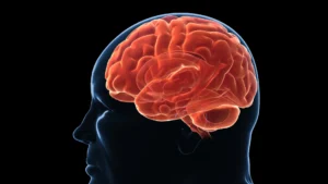

Barcelona, Spain – For decades, the medical community has categorized asthma strictly as a respiratory condition, focusing its clinical scope on the bronchial tubes, lung capacity, and airway inflammation. However, groundbreaking research presented this week at the European Respiratory Society (ERS) Congress in Barcelona suggests that the reach of asthma may extend far deeper into the human body—specifically, into the architecture of the brain.
A study conducted by researchers at the NIHR Biomedical Research Centre (BRC) in Bristol and City St George’s University of London has unveiled evidence that patients suffering from severe type-2 asthma exhibit subtle, yet measurable, differences in their brain structure compared to those whose asthma is well-managed. This discovery marks a potential paradigm shift in how clinicians perceive the systemic nature of severe chronic inflammatory diseases.
The Core Findings: A New Frontier in Asthma Research
The study, led by Dr. George Nava, an NIHR Academic Clinical Lecturer at the Bristol Academic Respiratory Unit, utilized advanced neuroimaging to peer into the neural pathways of asthma patients. By comparing 50 individuals diagnosed with severe type-2 asthma against 25 participants with mild-to-moderate, well-controlled asthma, the team identified distinct structural deviations in the former group.
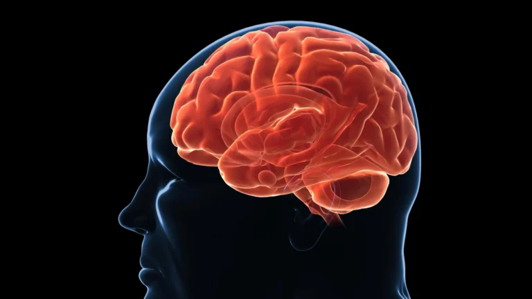
The research focused on "white matter," the brain’s complex communication network responsible for transmitting signals between various regions. Using a state-of-the-art MRI technique known as quasi-diffusion imaging (QDI), researchers were able to map the microscopic integrity of these neural pathways. The results were striking: patients with severe asthma demonstrated a significantly less organized white matter network. Furthermore, the study noted tangible variations in the volume of specific brain structures, most notably the hippocampus—the area associated with memory and spatial navigation—and the temporal lobes.
Most critically, the research team found a direct correlation between the degree of white matter disorganization and the levels of type-2 inflammation present in the patients. This suggests that the persistent, systemic inflammation characteristic of severe asthma may not be localized to the lungs but could be exerting a detrimental effect on central nervous system integrity.
Chronology: From Respiratory Focus to Neurological Investigation
The journey to this discovery began with the growing clinical suspicion that the impact of asthma was being underestimated.
- Initial Observations: For years, clinicians have noted that asthma patients often report cognitive and psychological comorbidities, including "brain fog," anxiety, and memory lapses. However, these symptoms were frequently attributed to secondary factors, such as sleep deprivation caused by nighttime breathing difficulties or the side effects of corticosteroid medications.
- Study Initiation: Recognizing the need for objective biological data, the team at the Bristol Academic Respiratory Unit and City St George’s University of London launched a comparative study to isolate the impact of the disease phenotype itself from external variables.
- Data Collection (2025–2026): Researchers recruited a cohort of 75 participants. Over the course of the study, patients underwent comprehensive lung function testing alongside high-resolution brain imaging.
- The ERS Congress Presentation (September 8, 2026): The findings were formally unveiled to the international respiratory community in Barcelona, triggering an immediate conversation regarding the systemic nature of inflammatory airway diseases.
Supporting Data: The Science of Inflammation
To understand why these brain changes occur, one must look at the nature of "Type-2" asthma. This phenotype is driven by an overactive immune response, leading to constant, low-grade systemic inflammation. While the lungs are the primary site of attack—characterized by eosinophilic inflammation, mucus production, and airway remodeling—the immune signaling molecules (cytokines) involved in this process circulate throughout the bloodstream.
The Role of QDI Imaging
The use of quasi-diffusion imaging (QDI) was instrumental in this study. Unlike standard MRI, which provides a macroscopic view of brain structure, QDI allows scientists to examine the microscopic architecture of white matter tracts. By measuring how water molecules diffuse along these pathways, researchers can determine the "health" or "organization" of the neural insulation (myelin). In the severe asthma cohort, the diffusion patterns indicated a degradation in the efficiency of these signal-carrying pathways, mirroring the type of neuro-inflammation seen in other chronic inflammatory conditions.
The Inflammation Correlation
The study’s most compelling data point is the positive correlation between biomarker levels (indicative of type-2 inflammation) and the extent of white matter disorganization. This suggests a dose-dependent relationship: the more severe the systemic inflammation, the greater the observable difference in brain architecture.
Official Responses and Expert Commentary
The presentation at the ERS Congress has drawn significant attention from global respiratory experts.
Dr. George Nava, lead researcher, summarized the gravity of the findings during his presentation:
"Asthma is usually thought of as a disease of the lungs, but growing evidence suggests its effects may extend well beyond the airways. We found measurable differences in brain structure in people with severe type-2 high asthma compared with people whose asthma was mild-moderate and well-controlled. While this study cannot prove that inflammation causes these changes, it does suggest there may be a biological link that deserves further investigation."
Professor James Dodd, Professor of Respiratory Medicine at the University of Bristol and Chief Investigator, emphasized the future-oriented nature of the research:
"One of the most important questions is whether these brain changes can be prevented or reversed. We are now pivoting our efforts to investigate whether these structural differences shift after patients begin biological therapies—treatments that specifically target the inflammation driving severe type-2 asthma."
Professor Apostolos Bossios, Head of the ERS Assembly on Airway Diseases, who was not involved in the study, offered a broader clinical perspective:
"We already know that asthma can have effects beyond the lungs, including impacts on memory, thinking, and wellbeing. Type-2 asthma is the most common phenotype, yet 5–10% of patients develop a severe form that is resistant to traditional therapy. This study provides a vital missing piece of the puzzle. It suggests that if we can suppress the systemic inflammation more effectively, we may be protecting more than just the patient’s breathing—we may be protecting their brain health."
Clinical Implications: A New Era for Asthma Management?
The implications of these findings are profound, potentially altering the standard of care for severe asthma patients in several ways:
1. Reassessing Treatment Urgency
If asthma is confirmed to have neuro-inflammatory consequences, the urgency for early and aggressive intervention becomes even higher. The current "step-up" approach to asthma treatment—where medications are increased only as symptoms worsen—might be insufficient if the goal is to prevent long-term neurological impact.
2. The Potential for Reversibility
The primary question currently facing the research team is the reversibility of these structural changes. If biological therapies—such as monoclonal antibodies that neutralize specific inflammatory cytokines—can "reset" the white matter organization, it would provide a powerful incentive for early adoption of advanced therapies.
3. Broadening the Diagnostic Scope
Physicians may soon need to incorporate cognitive health screenings into the routine care of severe asthma patients. Recognizing signs of cognitive decline or executive dysfunction could prompt earlier intervention or a shift toward more targeted, systemic anti-inflammatory treatments.
4. A Multi-Disciplinary Approach
This research signals a move away from the "siloed" medical model. Moving forward, the treatment of severe asthma may require a collaboration between pulmonologists, neurologists, and immunologists to manage the patient’s health in a holistic, systemic fashion.
Conclusion: A Call for Further Investigation
While the results presented in Barcelona are transformative, the research team is cautious, noting that the study establishes an association rather than definitive causation. The small sample size and the cross-sectional nature of the study mean that more extensive, longitudinal research is required.
As the scientific community digests these findings, the focus will undoubtedly shift to the "why" and "how." Is the brain damage a direct result of inflammatory molecules crossing the blood-brain barrier, or is it an indirect consequence of chronic hypoxia and sleep disturbance?
Whatever the mechanism, the message from the 2026 ERS Congress is clear: the patient experience of severe asthma is far more complex than a struggle for breath. By looking beyond the airways and into the mind, medicine is finally beginning to uncover the hidden costs of living with severe, chronic inflammation. The future of asthma treatment may well depend on our ability to heal the brain as effectively as we heal the lungs.