





For decades, the standard laboratory model for studying human blood vessels was remarkably, and perhaps dangerously, simplistic. Scientists relied on straight, uniform tubes to replicate the complex highways of the human circulatory system. While these models provided a baseline for fundamental research, they ignored the physiological reality that blood vessels are dynamic, tortuous, and highly irregular. In the human body, vessels bend, branch, narrow, and widen, creating intricate hemodynamic environments that play a decisive role in the onset and progression of vascular diseases.
Recognizing this critical disconnect between laboratory models and human biology, researchers at the Department of Biomedical Engineering at Texas A&M University have unveiled a groundbreaking advancement: a customizable, "vessel-chip" system. This innovation, which promises to redefine how scientists study vascular pathology and test pharmaceutical interventions, moves the field away from static, idealized designs toward living, architectural replicas of the human vasculature.
The Architecture of Life: A New Paradigm in Microfluidics
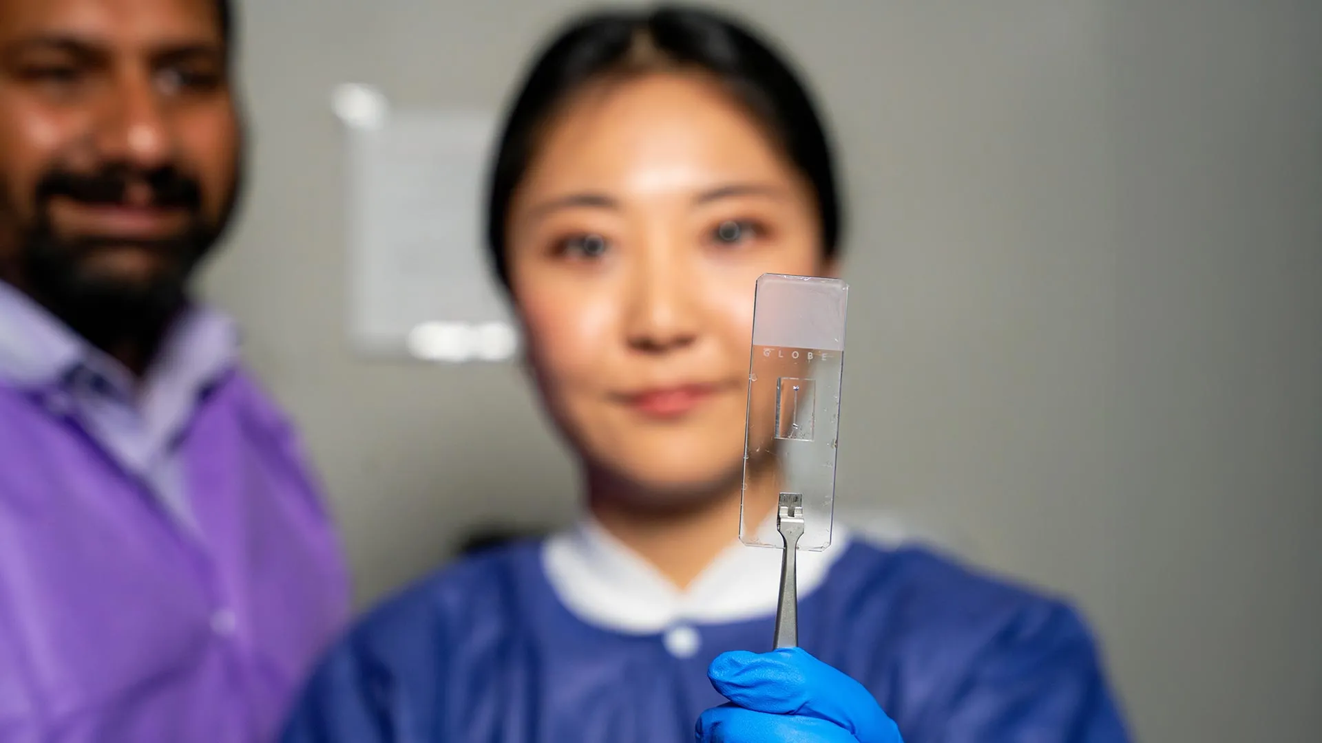
The core of this research, led by Jennifer Lee under the guidance of Dr. Abhishek Jain, lies in the development of microfluidic devices that mimic the structural complexity of human vessels at a microscopic scale. These chips are not merely plastic molds; they are sophisticated, bio-engineered environments that can be tailored to individual patient profiles, offering a robust, non-animal alternative for clinical modeling.
The research, which is set to be featured on the cover of the May 2025 issue of the prestigious journal Lab on a Chip, represents a significant leap forward in the "organs-on-a-chip" field. Unlike their predecessors, these new chips can replicate the specific geometries that characterize clinical conditions, such as aneurysms—where vessels expand dangerously—or stenosis, where they narrow and restrict blood flow.
The Physics of Flow and Shear Stress
The significance of these geometries cannot be overstated. According to Lee, the shape of the vessel directly dictates the behavior of the blood flowing through it. "There are branched vessels, or aneurysms that have sudden expansion, and then stenosis that restricts the vessel," Lee explains. "All these different types of vessels cause the blood flow pattern to be significantly changed, and the inside of the blood vessel is affected by the level of shear stress caused by these flow patterns. That’s what we wanted to model."
Shear stress—the frictional force exerted by blood flowing against the vessel wall—is a primary trigger for cellular responses. By successfully mimicking these varying stress environments, the Texas A&M team has created a platform where scientists can observe, in real-time, how cells react to the mechanical forces that lead to plaque buildup, clotting, and other vascular failures.
A Chronology of Innovation: From Straight Tubes to Complex Systems
The journey to this sophisticated vessel-chip was one of iterative progress within the Bioinspired Translational Microsystems Laboratory. The lab, directed by Dr. Abhishek Jain, an associate professor and the Barbara and Ralph Cox ’53 faculty fellow in biomedical engineering, has long been at the forefront of micro-physiological systems.
The Foundation: The Straight Vessel Era
A few years prior to Lee’s breakthrough, Dr. Tanmay Mathur, a former graduate student in the Jain lab, established the proof-of-concept for the straight vessel-chip. This initial model provided the essential technical framework: how to interface living cells with micro-scale fluidics and how to maintain cellular viability under constant flow. While the straight model was a necessary first step, it was limited in its ability to simulate the diverse, high-risk regions of the circulatory system where disease typically manifests.
The Expansion: Jennifer Lee’s Contribution
Jennifer Lee joined the lab as an undergraduate honors student, entering a field with which she had little prior experience. Her transition from a novice to the lead developer of an advanced, complex vessel-chip is a testament to the university’s commitment to undergraduate research. Under Dr. Jain’s mentorship, Lee pushed the boundaries of the existing technology, focusing on the fabrication techniques required to mold complex, non-uniform geometries into the microfluidic substrates. Her work successfully transitioned the lab’s focus from "simple tubes" to "complex conduits," bridging the gap between basic engineering and clinical application.
The Power of "Living" Chips: Implications for Medicine
The primary advantage of the vessel-chip system is that it is not a lifeless model; it is a living, breathing biological system. As Dr. Jain emphasizes, the ability to populate these structures with actual cellular and tissue material changes the scope of what is possible.
"We can now start learning about vascular disease in ways we’ve never been able to before," says Jain. "Not only can you make these structures complex, you can put actual cellular and tissue material inside them and make them living. These are the sites where vascular diseases tend to develop, so understanding them is critical."
Future-Proofing Drug Discovery
The implications for the pharmaceutical industry are profound. Current drug testing often relies on animal models that may not accurately predict human vascular responses, leading to high failure rates in clinical trials. By using human-derived cells in a vessel-chip that replicates a patient’s specific vascular architecture, researchers can conduct "clinical trials on a chip." This allows for the high-throughput testing of drugs, the evaluation of personalized medicine protocols, and the study of rare vascular conditions without the ethical and logistical constraints of animal research.
Moving Toward the Fourth Dimension
Despite the success of the current design, the research team is already looking toward the next frontier. Currently, the model focuses on endothelial cells—the specialized cells that line the interior surface of blood vessels. While this provides an excellent look at the vessel-blood interface, the team is working on incorporating additional cell types, such as smooth muscle cells and pericytes, to recreate the multi-layered structure of a mature vessel.
Dr. Jain describes this evolution as the "fourth dimensionality of organs-on-a-chip." In this paradigm, researchers move beyond the static study of cell types and flow patterns to explore the dynamic interaction between various tissues and blood flow within complex architectural states. This holistic approach is essential for understanding diseases like atherosclerosis, which involves not just the lining of the vessel, but the entire wall structure.
Beyond the Bench: Developing Future Scientists
The story of this research is as much about the human element as it is about engineering. For Jennifer Lee, the project served as an immersive educational experience that extended far beyond the technical aspects of microfluidics. Joining the lab as an undergraduate provided her with a unique mentorship structure that fostered critical thinking, resilience, and interdisciplinary collaboration.
"It’s such a good environment to interact with not only peers but also graduate students and postdoctoral researchers," Lee says. "You’re able to learn teamwork and communication, work ethic, and just trying different things out. I think it’s such a valuable experience that students have available."
Dr. Jain underscores the value of the university’s fast-track master’s program, which enabled Lee to take her undergraduate curiosity and translate it into a high-impact, published scientific outcome. "Jennifer demonstrated perseverance, curiosity, and creativity," Jain notes. "Our program enables students to take on high-impact, high-risk research and not just do a science project, but take it all the way to its outcome and get it published."
Sustaining the Vision: Institutional and Federal Support
The scale and scope of this research have been made possible by a broad coalition of support from national and federal agencies. This level of interest highlights the importance of the work for both national health and space exploration. The research received funding and support from:
- The U.S. Army Medical Research Program: Recognizing the potential for military trauma and vascular injury care.
- NASA: Investigating how vascular health is impacted by microgravity, a key area of study for long-duration spaceflight.
- The Biomedical Advanced Research and Development Authority (BARDA): Focused on medical countermeasures for public health threats.
- The National Institutes of Health (NIH) and the FDA: Supporting the move toward more reliable, human-relevant preclinical models.
- The National Science Foundation (NSF): Providing the fundamental research funding necessary for innovative breakthroughs.
- Texas A&M University Office of Innovation Translational Investment Funds: Ensuring that university research is translated into viable, real-world solutions.
Conclusion: A New Horizon for Vascular Health
As the field of biomedical engineering continues to advance, the work being done in Dr. Jain’s lab serves as a beacon for what is possible when engineering precision meets biological complexity. By moving beyond the "straight tube" mentality, the Texas A&M team has opened a window into the nuanced, high-stakes world of human blood flow.
The vessel-chip system is more than a tool; it is a platform for discovery that promises to accelerate our understanding of vascular disease and improve the lives of patients worldwide. As Lee’s research reaches the cover of Lab on a Chip this May, it stands as a testament to the power of student-led innovation and the critical importance of realistic, bio-inspired models in the modern era of medicine.