







For millions struggling with alcohol use disorder (AUD), the cycle of addiction is not a simple pursuit of pleasure, but a desperate, repetitive flight from pain. While society often views addiction through the lens of hedonistic indulgence, a groundbreaking study published August 5, 2025, in Biological Psychiatry: Global Open Science suggests that the biological reality is far more somber. Researchers at Scripps Research have identified a specific neurological “switch” that traps individuals in a cycle of relapse, driven not by the desire to get high, but by the physiological necessity to escape the agony of withdrawal.
The Architecture of Escape: Main Facts and Findings
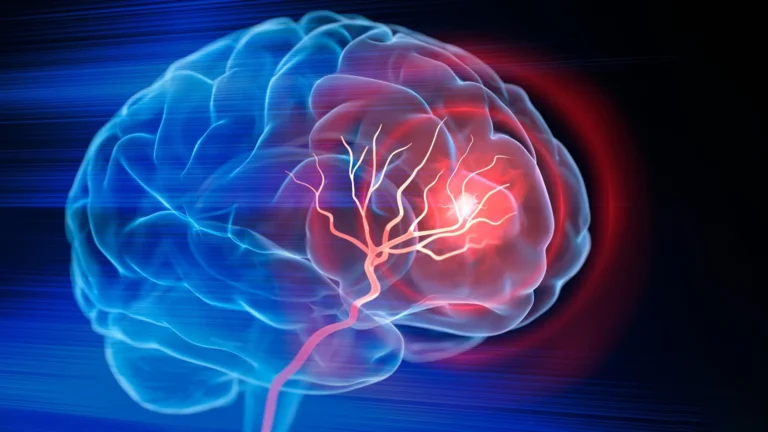
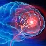
At the heart of this study is the paraventricular nucleus of the thalamus (PVT), a small but critical region of the brain involved in the regulation of stress and arousal. By observing rat models, the research team discovered that the PVT serves as the primary neural substrate where the brain “learns” that alcohol is a remedy for the physical and emotional distress of withdrawal.
The study indicates that when an animal experiences the relief provided by alcohol during a state of withdrawal, the PVT becomes hyper-active. This activity creates a powerful, persistent association between environmental cues—the sights, sounds, or locations where drinking occurred—and the cessation of withdrawal symptoms. This process, known in neuroscience as negative reinforcement, effectively "locks in" the addictive behavior, making it incredibly difficult to extinguish even when the subject is faced with negative consequences or punishing stimuli.
A Chronology of Discovery: From Pleasure to Compulsion
To understand how addiction evolves, one must look at the progression of the brain’s relationship with alcohol. The research team, led by senior authors Friedbert Weiss and Hermina Nedelescu, built upon their previous 2022 research, which mapped the behavioral trajectory of AUD.
Phase 1: The Initial Reward
In the early stages of exposure, alcohol consumption is largely driven by positive reinforcement—the “high.” The brain’s reward centers are activated, creating a dopaminergic loop that encourages the subject to seek out alcohol to repeat the pleasurable experience.
Phase 2: The Withdrawal Threshold
As cycles of heavy drinking and subsequent abstinence continue, the brain undergoes neurochemical adaptations. The absence of alcohol no longer merely represents a return to baseline; it triggers a “negative hedonic state.” This state is characterized by profound anxiety, physical tremors, and extreme physiological stress.
Phase 3: The Shift to Negative Reinforcement
The current 2025 study highlights the pivotal moment when the motivation for drinking shifts. Once the subject discovers that alcohol acts as a chemical anesthetic for the pain of withdrawal, the brain reconfigures its priorities. The PVT becomes the command center for this new motivation. The researchers observed that even when alcohol was no longer producing a “high,” the subjects would work tirelessly—persisting through punishing conditions—solely to reach the relief associated with their environment.
Mapping the Neural Landscape: Supporting Data
To pinpoint the PVT as the culprit, the research team utilized advanced whole-brain imaging techniques. By comparing rats that had developed withdrawal-linked drinking habits against three distinct control groups, the team was able to isolate cellular activity patterns.
The data revealed a striking correlation: in every subject that had undergone withdrawal-related learning, the PVT “lit up” when exposed to cues previously associated with the relief of withdrawal. This suggests that the PVT is not just a passive bystander but an active, necessary component of the neural circuitry that sustains addiction.
The team’s methodology was rigorous, ensuring that the observed neural activity was specific to the relief of the “negative hedonic state” rather than general stress or the presence of alcohol alone. By stripping away the variables, they confirmed that the PVT is the specific locus where the brain encodes the concept of “relief.”
Official Perspectives: Insights from the Researchers
The findings from the Scripps team challenge long-standing assumptions about the nature of substance use disorders. Dr. Friedbert Weiss, a professor of neuroscience at Scripps Research, emphasizes that this research is a necessary correction to the popular understanding of addiction.
"What makes addiction so hard to break is that people aren’t simply chasing a high," Dr. Weiss states. "They’re also trying to get rid of powerful negative states, like the stress and anxiety of withdrawal. This work shows us which brain systems are responsible for locking in that kind of learning, and why it can make relapse so persistent."
Co-senior author Hermina Nedelescu adds that the discovery of the PVT’s role provides a tangible target for future medical interventions. "This brain region just lit up in every rat that had gone through withdrawal-related learning," Dr. Nedelescu explains. "It shows us which circuits are recruited when the brain links alcohol with relief from stress—and that could be a game-changer in how we think about relapse."
Implications: A New Horizon for Treatment and Beyond
The implications of identifying the PVT as a central hub for negative reinforcement extend far beyond alcohol use disorder. The researchers suggest that the mechanisms identified in this study are universal features of the human brain’s survival systems.
Addressing Substance Use Disorders (SUDs)
By isolating the neural pathways involved in negative reinforcement, scientists may be able to develop pharmacological interventions that “dampen” the PVT’s reactivity. If a treatment could block the brain from reinforcing the connection between environmental cues and the relief of withdrawal, it could theoretically decouple the act of drinking from the drive to escape stress. This would empower patients to engage in recovery without the constant, overwhelming urge to seek relief.
Implications for Mental Health
The study also suggests a broader application for anxiety disorders, fear-conditioning, and traumatic avoidance learning. In many of these conditions, the patient acts to escape a perceived threat or an internal state of distress. If the PVT is the universal site for this type of “escape learning,” therapies targeting this region could offer new avenues for treating generalized anxiety, panic disorders, and PTSD.
The Road Ahead: Future Research Directions
The team at Scripps Research is not stopping here. Their next phase of research will focus on two key areas:
- Biological Inclusivity: Expanding the study to include female subjects to determine if there are sex-based differences in how the PVT regulates withdrawal-related learning.
- Molecular Targeting: Investigating the specific neurochemicals released within the PVT during these moments of relief. By identifying the unique receptors and molecules at play, researchers hope to provide pharmaceutical companies with a blueprint for drug development.
Conclusion: Reframing the Struggle
The research published in Biological Psychiatry: Global Open Science serves as a sobering reminder of the biological chains that bind those with alcohol use disorder. It moves the conversation away from moral judgment and toward a sophisticated understanding of brain circuitry.
As the scientific community continues to map the geography of the brain, the identification of the PVT as a core component of addiction provides a new sense of hope. By acknowledging that addiction is often a desperate, learned response to internal suffering, we move closer to creating treatments that are not only more effective but also more compassionate. The work performed by Weiss, Nedelescu, and their colleagues suggests that the key to breaking the cycle of addiction may lie in the very region of the brain that once helped the individual survive the pain of withdrawal.
This study was supported by funding from the National Institutes of Health (NIH), including grants T32AA007456, K01 DA054449, R01 AA027555, and R01 AA023183. The research team included contributors from Scripps Research and MBF Bioscience.