





For decades, the standard laboratory approach to studying the human circulatory system has relied on a fundamental simplification: the straight, uniform tube. While these models have served as the bedrock of vascular biology, they represent a stark departure from the anatomical reality of the human body. In nature, blood vessels are dynamic, branching networks characterized by intricate geometry—bends, sudden narrowings, and expansive aneurysms. These architectural complexities are not merely incidental; they are the primary sites where vascular diseases, such as atherosclerosis and thrombosis, take root.
Now, a team of researchers in the Department of Biomedical Engineering at Texas A&M University is challenging this status quo. By developing a customizable "vessel-chip" system, they have bridged the gap between basic laboratory research and the chaotic, high-stakes environment of human hemodynamics.
The Problem with Simplification: Why Geometry Matters
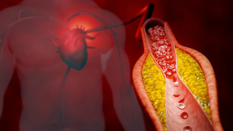
To understand the significance of this breakthrough, one must first understand the fluid dynamics of the human body. Blood flow is rarely laminar or uniform. When a vessel branches or narrows, it creates shifts in "shear stress"—the frictional force exerted by blood flowing against the vessel walls.
"There are branched vessels, or aneurysms that have sudden expansion, and then stenosis that restricts the vessel," explains Jennifer Lee, a master’s student in biomedical engineering who spearheaded the development of this new platform. "All these different types of vessels cause the blood flow pattern to be significantly changed, and the inside of the blood vessel is affected by the level of shear stress caused by these flow patterns. That’s what we wanted to model."
Traditional models, which treat vessels as standardized straight conduits, fail to capture the mechanical cues that tell cells to become inflamed, damaged, or diseased. By failing to replicate the "bends and branches" of the human anatomy, previous research may have missed critical insights into how vascular pathologies actually initiate.
A Chronology of Innovation: From Straight Tubes to Complex Systems
The journey to the current vessel-chip iteration began years ago within the Bioinspired Translational Microsystems Laboratory, led by Dr. Abhishek Jain.
The trajectory of this research reflects a deliberate, iterative process:
- The Foundation (Pre-2020s): The lab began by establishing the viability of microfluidic "organ-on-a-chip" technology. This technology allows scientists to culture human cells within tiny, clear devices that mimic the micro-environment of a living organ.
- The Initial Breakthrough: Dr. Tanmay Mathur, a former graduate student in the lab, successfully engineered a "straight vessel-chip." This provided the essential proof-of-concept that human vascular cells could be successfully cultured in a microfluidic device and subjected to controlled flow.
- The Architectural Expansion (2023–2024): Building upon Mathur’s work, Jennifer Lee undertook the challenge of introducing complex geometry. Her design allows for the customization of vessel shapes, moving beyond the linear paradigm to incorporate the diverse architectures found in the human body.
- Peer Validation (2025): The culmination of this effort was the publication of the research in the journal Lab on a Chip. The study has garnered significant professional attention, earning a coveted cover feature for the May 2025 issue.
Technical Infrastructure: The "Vessel-on-a-Chip"
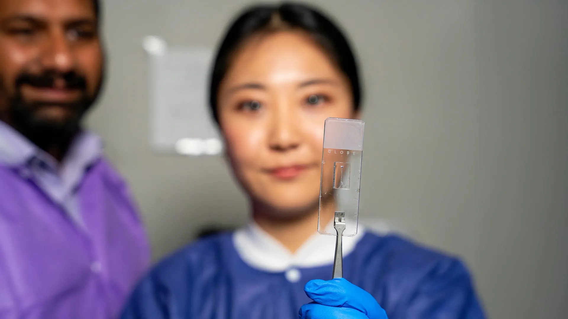
The device itself is a triumph of bioengineering. Microfluidic devices are fabricated with precision to replicate the scale of human capillaries and larger vessels. These chips are not just passive models; they are "living" systems.
Researchers seed the interior of these channels with endothelial cells—the delicate, specialized cells that line our blood vessels. By controlling the flow of nutrient-rich media through these chips, scientists can simulate the physiological conditions of the human heart and circulatory system.
Crucially, these devices are customizable. They can be tailored to replicate the specific vascular architecture of individual patients, opening the door for a new era of "personalized medicine." If a patient has a specific type of stenosis (narrowing), a chip can be designed to mimic that exact geometry to test how various pharmaceutical interventions might perform in that specific, high-risk environment.
The Vision of Dr. Abhishek Jain: The "Fourth Dimension"
Dr. Abhishek Jain, an associate professor and the Barbara and Ralph Cox ’53 faculty fellow, views this development as a pivotal shift in the field of micro-physiological systems. He describes the move toward complex vessel architecture as entering the "fourth dimension" of organ-on-a-chip research.
"We can now start learning about vascular disease in ways we’ve never been able to before," Dr. Jain says. "Not only can you make these structures complex, you can put actual cellular and tissue material inside them and make them living. These are the sites where vascular diseases tend to develop, so understanding them is critical."
By moving beyond the cell-and-flow model, Jain’s team is now looking toward the integration of multi-tissue systems. Future iterations of the vessel-chip will incorporate diverse cell types, such as smooth muscle cells or immune cells, to create a more comprehensive "vascular ecosystem." This allows researchers to study how different tissues interact with each other and with flowing blood under stress, providing a holistic view of disease progression that simply cannot be observed in traditional, static cell cultures.
Academic Growth: Mentorship and the Fast-Track Path
The success of this research is also a testament to the effectiveness of the Texas A&M biomedical engineering curriculum. Jennifer Lee’s path from an undergraduate honors student to a published lead researcher underscores the department’s commitment to "high-impact, high-risk" research.
When Lee first joined the lab, she had little experience with the intricacies of microfluidics. However, the lab’s collaborative culture—which integrates undergraduates, master’s students, and postdoctoral fellows—provided a scaffold for her rapid development.
"Jennifer demonstrated perseverance, curiosity, and creativity and started taking up research projects very quickly," Dr. Jain notes. "Our fast-track program enables students like Jennifer to take on sort of high-impact, high-risk research and not just do a science project, but take it all the way to its outcome and get it published."
For Lee, the experience was about more than just data collection. It was about developing the professional competencies required in the modern scientific landscape: communication, interdisciplinary teamwork, and the resilience to troubleshoot complex hardware failures. "It’s such a good environment to interact with not only peers but also graduate students and postdoctoral researchers," Lee reflects. "You’re able to learn teamwork and communication, work ethic, and just trying different things out."
Implications for Future Medical Science
The broader implications of the Texas A&M vessel-chip are profound.
1. Reducing Reliance on Animal Models
Animal testing has long been the standard for preclinical drug trials, yet animals often do not accurately predict how a drug will interact with the human circulatory system. The vessel-chip offers a human-centric, ethical alternative that can provide high-fidelity data on drug efficacy and toxicity without the ethical and biological limitations of animal subjects.
2. Accelerating Drug Discovery
By using these chips, pharmaceutical companies can perform high-throughput screening of drug candidates. Because the chips can be produced in batches and subjected to automated, high-precision flow, researchers can test thousands of compounds to see how they affect vascular health under various flow conditions.
3. Personalized Medicine
Perhaps the most exciting frontier is the use of patient-derived cells. By taking a small sample of a patient’s own cells and placing them in a chip modeled after their own vascular anatomy, doctors could potentially test which blood-thinning medication or intervention will be most effective for that specific individual before ever administering the drug to the patient.
A Collaborative Endeavor
The magnitude of this research has attracted the attention and support of major national and international agencies. The project’s success is rooted in the financial and technical backing of the U.S. Army Medical Research Program, NASA, the Biomedical Advanced Research and Development Authority (BARDA), the National Institutes of Health (NIH), the U.S. Food and Drug Administration (FDA), and the National Science Foundation (NSF).
Additionally, the Texas A&M University Office of Innovation Translational Investment Funds provided the crucial support necessary to move the technology from the benchtop toward potential clinical translation.
Conclusion: The Path Forward
As the team continues to refine their vessel-chip technology, the focus remains on pushing the boundaries of what is possible. By adding more cell types and refining the geometric complexity of their chips, the researchers at Texas A&M are not just building models—they are building a bridge to a future where vascular disease can be understood, diagnosed, and treated with unprecedented precision.
The shift from the "straight tube" to the complex, branching reality of the human vessel is more than just a design change; it is a fundamental shift in how we perceive the mechanics of life. With the publication of their work in Lab on a Chip, the team has set a new standard for the field, ensuring that the next generation of vascular research will be as complex, dynamic, and intricate as the human body itself.