








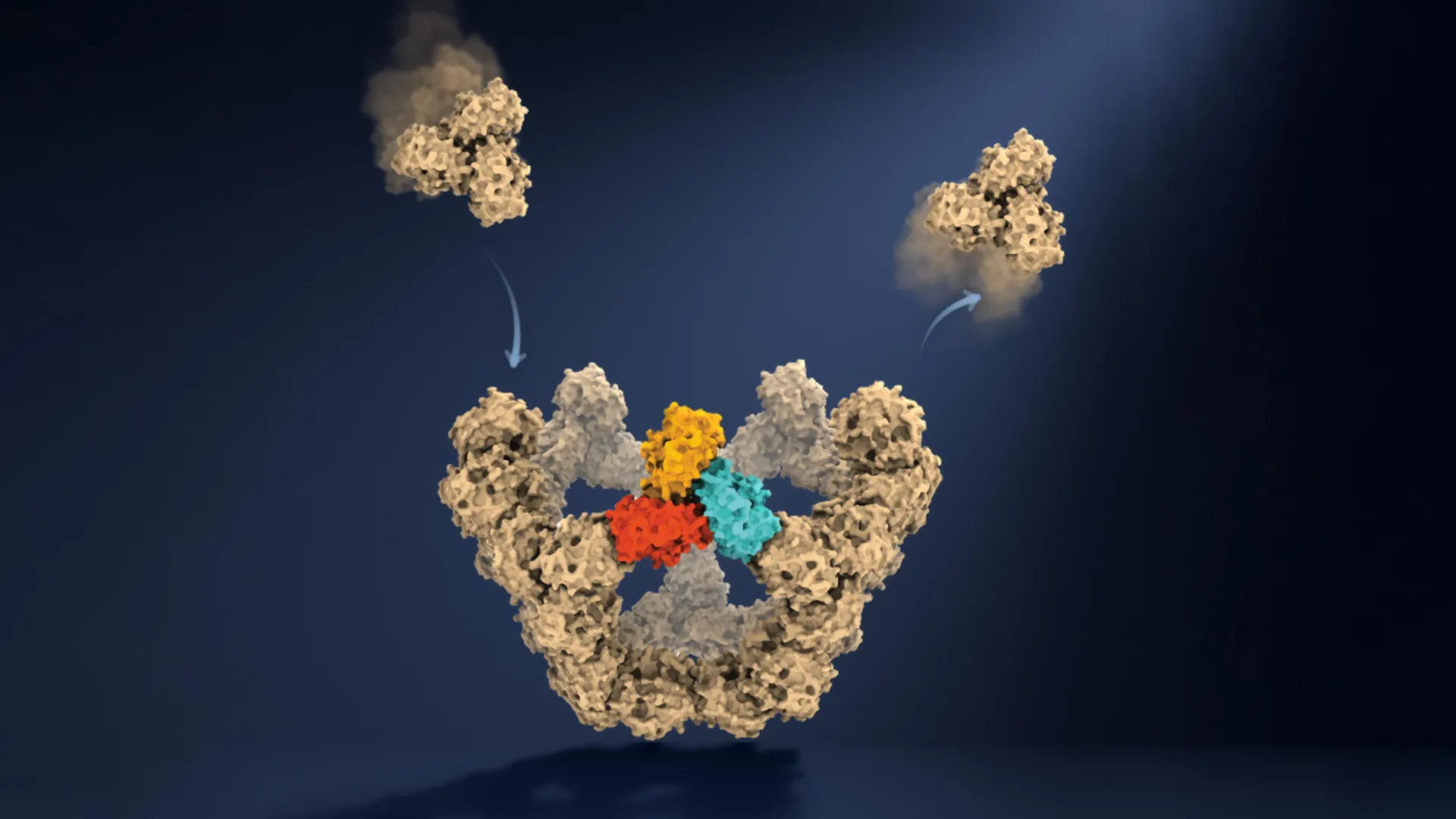
In the microscopic theater of biology, few processes are as consequential—or as mysterious—as the formation of a virus. For decades, the spontaneous assembly of viral shells, known as capsids, has been the subject of theoretical modeling and fragmented snapshots. Now, researchers at the University of Oxford have achieved what was once considered impossible: they have watched, in real-time and molecule-by-molecule, as virus-like particles construct themselves from the ground up.
The study, published in the journal Nature, reveals that the "simple" rules governing viral assembly are a masterclass in molecular efficiency. By observing the process as it unfolds, the Oxford team has effectively demystified how complex biological structures form with near-perfect reliability, paving the way for a new generation of precision medicine, antiviral therapies, and advanced vaccine technology.
The Main Facts: Cracking the Viral Blueprint
Viruses are, in essence, genetic masterminds encased in sophisticated protein armor. To replicate, they must shield their genetic material within a capsid, a protective shell composed of protein building blocks that can range from a few dozen to several thousand units. Despite the staggering complexity of these structures, they assemble without the aid of internal machinery or an external blueprint.
The Oxford research confirms that this process is not a chaotic collision of parts, but a refined, self-correcting pathway. The team discovered that viral assembly functions similarly to navigating a high-stakes maze. Proteins form weak, tentative connections that are designed to fail; if a configuration is incorrect, the bond breaks, allowing the components to detach and try again. This "trial-and-error" mechanism ensures that only the most robust, geometrically sound configurations persist. Once a threshold of stability is reached, the structure "locks in," effectively narrowing the astronomical number of potential assembly routes into a streamlined, high-speed path toward completion.
Chronology: A New Lens on Molecular Dynamics
The journey to this discovery began with the limitation of traditional observation methods. For years, scientists relied on techniques like cryo-electron microscopy, which provided high-resolution "snapshots" of the starting proteins and the finished viral shell. However, the intermediate states—the fleeting, unstable configurations that bridge the gap between individual proteins and a fully formed capsid—remained elusive. These intermediates are often short-lived and present in such low concentrations that they vanish before they can be captured.
To bypass this, the Oxford team turned to mass photometry. This sophisticated technology, pioneered at the University of Oxford, measures the light scattered by individual molecules to determine their precise mass. By refining this technique and developing a specialized confinement method, the team was able to keep individual, growing viral particles under continuous observation.
Over a series of experiments, researchers tracked the assembly of an engineered 60-protein virus-like particle. The process was recorded with such granular detail that the researchers could effectively watch the assembly like a video, tracking the mass of the particle as each individual building block was added to the structure. This chronological data allowed the team to map the assembly trajectory for the first time, identifying the exact moments where individual units transition from unstable, random attachments to permanent, structural components.
Supporting Data: The Pentagonal Milestone
A pivotal finding in the study involves the concept of "nucleation"—the initial, and often most arduous, stage of assembly. The data indicates that the assembly process is not linear; rather, it is characterized by specific "checkpoints."
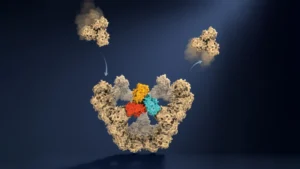
The most critical of these occurs when five protein building blocks connect to form a closed pentagonal ring. This structure serves as the first highly stable "anchor" in the construction process. Before this pentagonal ring forms, the system is prone to frequent disassemblies. However, once this foundation is laid, the thermodynamics of the system shift dramatically. Each subsequent stage of growth requires fewer additional building blocks to stabilize, leading to an exponential increase in assembly speed.
The Oxford team’s quantitative data provides a clear explanation for this phenomenon:
- Early Phase: High trial-and-error frequency; weak bonds; low stability.
- Nucleation Milestone: Formation of the pentagonal ring; the first "locked-in" structure.
- Growth Phase: Rapid, accelerated addition of remaining subunits, guided by the stability of the established framework.
This model provides a clear, evidence-based answer to the long-standing question of how a system with thousands of possible wrong turns consistently arrives at the same, highly organized destination.
Official Responses: From the Lab Bench to the Future
The research team, comprised of experts from the Department of Chemistry and the Kavli Institute for Nanoscience Discovery, emphasized the transformative nature of their findings.
"A virus has to solve an extraordinary construction problem," explains co-lead author Dr. Roi Asor. "Its components somehow have to find the right arrangement among a huge number of possibilities, without a blueprint or machinery directing the process. We can now watch what happens molecule by molecule and see the physical rules that make it possible."
The ability to move from theoretical models to direct observation has changed the researchers’ fundamental understanding of the system. Professor Philipp Kukura, co-lead author, noted, "Until now, much of our understanding of how these structures assemble has had to be reconstructed from snapshots or theoretical models. Being able to both quantify the underlying interactions and follow one particle as it grows changes that. We can see the important intermediate structures appear and deduce a detailed model of the construction process."
For PhD student and co-first author Dan Loewenthal, the excitement lies in the simplicity of the approach: "This method lets us study the assembly process directly; we just take a video!"
Implications: A New Frontier in Medicine and Beyond
The implications of this discovery extend far beyond the study of viral architecture. By deciphering the "rules of engagement" for viral self-assembly, scientists now have a targetable, logical pathway for intervention.
1. Disrupting Viral Replication
Current antiviral drugs often target the virus’s enzymes or surface receptors. However, by understanding the molecular mechanics of how a capsid is built, researchers could design "assembly-inhibitor" drugs. These compounds would act as molecular wrenches, targeting the specific intermediate stages of assembly—such as the formation of the pentagonal ring—to prevent the virus from ever completing its protective shell. If the shell cannot be built, the viral genome remains vulnerable to degradation, effectively neutralizing the infection.
2. Next-Generation Vaccines
The study also provides a roadmap for bioengineering. By mastering the self-assembly of virus-like particles, researchers can improve the design of vaccines that use these particles to mimic the appearance of a pathogen without the risk of causing disease. Understanding how to "steer" the assembly process allows for higher yields and more consistent, uniform particles, which are essential for effective immunization.
3. Broad Biological Applications
The principles uncovered at Oxford are universal. Self-assembly is a fundamental biological process, responsible for everything from the formation of cellular skeletons (the cytoskeleton) to the creation of complex protein compartments that govern metabolic processes. The ability to watch these processes in real-time opens the door to understanding how errors in self-assembly contribute to diseases, including neurodegenerative disorders where proteins aggregate incorrectly.
"The ability to understand these processes at the molecular level could have important applications in vaccine development and antivirals that disrupt viral assembly," says co-author Dr. Jack Tan of the MRC Weatherall Institute of Molecular Medicine.
In conclusion, the Oxford study represents a paradigm shift in structural biology. By turning the "black box" of viral construction into a transparent, observable process, the team has not only solved a decades-old puzzle but has also provided the tools necessary to combat some of the most persistent challenges in modern medicine. As we continue to refine our ability to observe the microscopic world, we move closer to a future where we can out-engineer the viruses that have long plagued human health.