










For decades, the mammogram has stood as the cornerstone of breast health. Every October, during Breast Cancer Awareness Month, the focus sharpens on National Mammography Day, a time dedicated to reminding the public that early detection is not merely a clinical recommendation—it is a life-altering strategy. By identifying malignancies before they become palpable or symptomatic, screening provides a critical window of opportunity, potentially shifting the trajectory of a diagnosis from a complex, late-stage challenge to a manageable, early-stage intervention.
However, as medical technology advances, our understanding of breast cancer screening has become more nuanced. It is no longer just about the X-ray; it is about understanding the limitations of the technology, the impact of biological variables like breast density, and the emerging frontier of "cancer interception."
The Core Facts: Why Screening Remains the Gold Standard
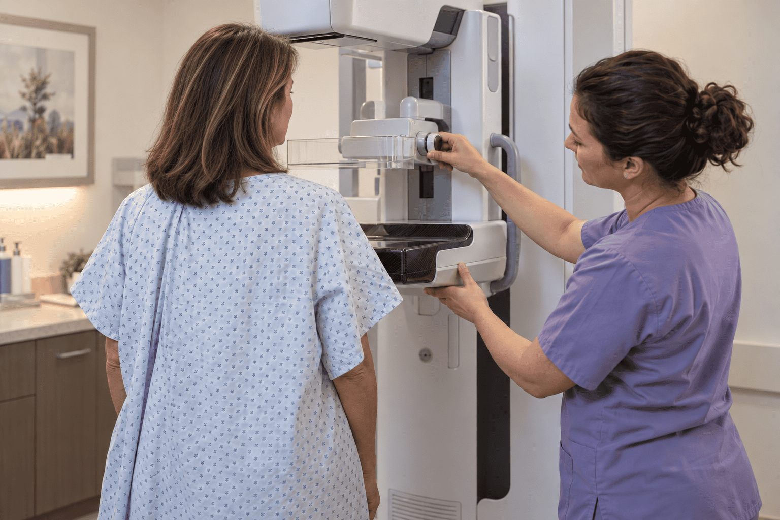
At its simplest, a mammogram is a specialized X-ray designed to capture detailed images of breast tissue. Its primary utility lies in its ability to detect microcalcifications or small, structural anomalies that are far too minute to be detected during a physical self-exam.
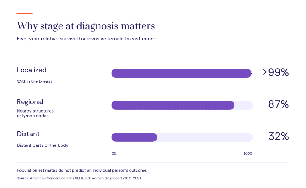
The rationale for regular screening is rooted in the biology of progression. When a cancer is localized—meaning it remains confined to the breast tissue where it originated—the survival outcomes are statistically favorable. According to current data from the Surveillance, Epidemiology, and End Results (SEER) program, the five-year relative survival rate for localized female breast cancer exceeds 99%. In contrast, that number drops to 87% for regional disease and falls to roughly 32% for distant disease, where the cancer has metastasized to other organs.

While these population-level statistics are not predictive of any single individual’s outcome, they offer a clear, irrefutable argument: the stage at which a tumor is identified is the single most significant factor in determining the spectrum of treatment options available.
A Chronology of Detection and Diagnosis
To understand the patient experience, one must look at the timeline of the diagnostic process.

- The Screening Baseline: A routine screening mammogram is performed on asymptomatic patients to establish a baseline of breast health.
- The "Callback" Phase: Approximately 10% of patients are asked to return for further imaging. It is crucial for patients to understand that a "callback" is not a cancer diagnosis. It is often a request for a clearer image, perhaps using a different angle or compression to rule out shadows, overlapping tissue, or benign cysts.
- Diagnostic Investigation: If the secondary imaging shows an area of concern, clinicians move to diagnostic tools. This may involve a targeted ultrasound to determine if a mass is fluid-filled (a cyst) or solid.
- Tissue Sampling: If the imaging remains suspicious, a biopsy is performed. A small sample of tissue is extracted and analyzed by a pathologist to determine if the cells are benign, precancerous, or malignant.
This systematic approach is designed to balance the need for vigilance with the reality that most "abnormal" findings on a primary mammogram are, in fact, benign.
Supporting Data: The Variables of Complexity
While the mammogram is a powerful tool, it is not infallible. The effectiveness of the scan is often dictated by individual biological factors, most notably breast density.
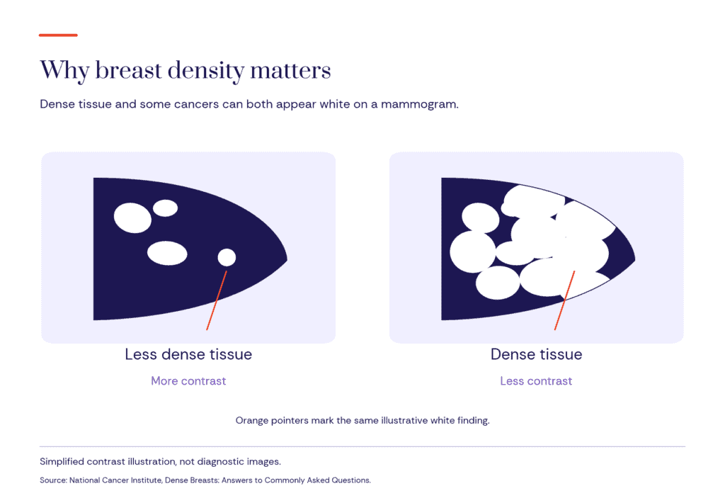
Nearly half of women over the age of 40 possess dense breast tissue—a term used to describe breasts that have more fibrous and glandular tissue compared to fatty tissue. On a mammogram, both dense tissue and tumors appear as white areas. This "white-on-white" contrast issue makes it significantly harder for radiologists to spot early-stage malignancies. Furthermore, medical research has consistently linked high breast density to an independent increase in breast cancer risk, making it a double-edged sword for many patients.
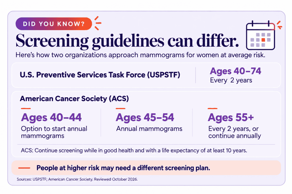
Because no single screening test is perfect, the medical community emphasizes the "trade-offs." Mammograms can result in false-positive results—which can lead to unnecessary anxiety and invasive procedures—and, in rare cases, they may miss cancers that are obscured by dense tissue. Additionally, there is the phenomenon of "overdiagnosis," where a test detects a slow-growing cancer that might never have caused health problems in the patient’s lifetime. These factors underscore why screening guidelines are not static; they are periodically updated by organizations like the American Cancer Society and the U.S. Preventive Services Task Force as new evidence emerges.

The Shift Toward "Cancer Interception"
Perhaps the most exciting development in the field is a shift in philosophy. Traditionally, screening was reactive: we looked for a tumor that had already formed. Today, leading researchers are asking: Can we stop the cancer before the tumor exists?
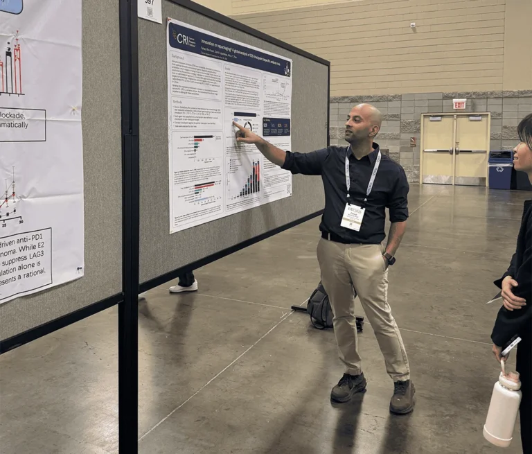
This emerging field, known as "cancer interception," seeks to identify the biological, genetic, and immunological "red flags" that precede the development of a tumor. Scientists are currently analyzing the tumor microenvironment—the ecosystem of cells, blood vessels, and nutrients that surround a developing cancer—to see if they can identify early immune system responses.
The Cancer Research Institute (CRI) is at the forefront of this movement. By investing in the Clinic and Laboratory Integration Program (CLIP), researchers are focusing on immune-based interventions. The goal is to move beyond simply "catching" cancer and toward a future where we can "intercept" the disease process, potentially preventing it from ever reaching the clinical stage.
Clinical Implications: A Personalized Approach
For the patient, the implications of these advancements are profound. The goal of modern medicine is to move toward personalized screening and treatment plans. This means that a 45-year-old with a family history of breast cancer and high breast density might have a very different screening schedule—incorporating MRIs or contrast-enhanced mammography—than a 45-year-old with low-density tissue and no family history.

Treatment today is also far less "one-size-fits-all." When cancer is caught early, the patient is often spared the most aggressive interventions. We are seeing a decrease in the necessity for radical mastectomies and intensive chemotherapy, replaced by targeted therapies, hormonal treatments, and breast-conserving surgeries. This shift is a direct result of earlier detection, which allows for smaller, less aggressive tumor management.
Official Guidance and Looking Ahead
As we reflect on National Mammography Day, the consensus among medical professionals remains clear: screening is the best defensive strategy we currently have. It is not about a guarantee of perfect health, but about maximizing the tools we have to secure the best possible outcomes.

However, the future is bright. As our understanding of the molecular origins of cancer grows, so too will our ability to detect disease with greater precision. The integration of artificial intelligence in image analysis is already helping radiologists identify patterns invisible to the human eye, and the advancements in liquid biopsies—blood tests that look for circulating tumor DNA—may one day complement the mammogram as a standard part of breast health surveillance.
For now, the message to the public is one of empowerment. Patients are encouraged to have open, honest conversations with their healthcare providers about their personal risk factors, their breast density, and the latest screening recommendations.
The window of opportunity in breast cancer care is widening. By staying informed, adhering to personalized screening schedules, and supporting the research that is driving the next generation of diagnostics, we can move closer to a world where a breast cancer diagnosis is not an end, but a manageable hurdle in a long and healthy life.
Sources for Further Reading
- American Cancer Society (2026): Survival Rates for Breast Cancer. An in-depth look at how stage-at-diagnosis affects long-term patient outcomes.
- Cleveland Clinic (2026): What is a Mammogram? A comprehensive guide on the mechanics and purpose of screening X-rays.
- Mayo Clinic: Understanding Your Mammogram Report. A resource for patients to navigate the terminology of radiology results and follow-up procedures.
- Cancer Research Institute (2027): Clinic and Laboratory Integration Program (CLIP). Information on current initiatives in immune-based cancer prevention.