


In a significant leap forward for molecular biology and pharmacology, a research team at the University of Maryland, Baltimore County (UMBC) has decoded a fundamental mystery of how enteroviruses hijack human cells to replicate. The study, recently published in the prestigious journal Nature Communications, details the precise molecular mechanics that viruses—ranging from the common cold to the more debilitating polio, myocarditis, and encephalitis—utilize to seize control of host cellular machinery.
By visualizing the interaction between viral RNA and specialized proteins at an atomic level, the UMBC researchers have not only clarified a decades-old scientific debate but have also unveiled a promising new target for the development of broad-spectrum antiviral medications. This discovery marks a pivotal shift in how scientists might approach the treatment of viral infections, moving away from single-pathogen therapies toward drugs capable of neutralizing entire families of viruses.
The Architecture of Invasion: Understanding Enteroviruses
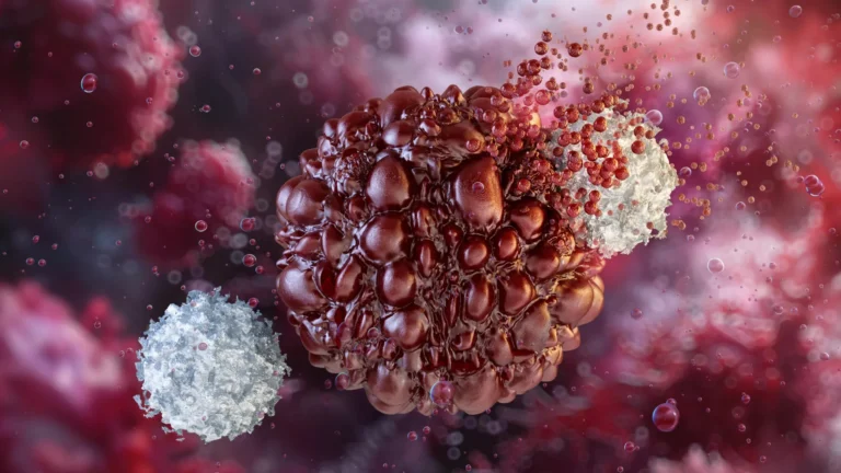
Enteroviruses are a genus of small, positive-sense, single-stranded RNA viruses. Despite their relatively simple genetic structure, they are remarkably efficient pathogens. Their ability to cause a wide spectrum of human illnesses—from mild respiratory symptoms to life-threatening neurological and cardiac conditions—stems from their sophisticated evolutionary strategies.
At the heart of the UMBC study is the "cloverleaf" structure, a highly specific folded region of the viral RNA genome. As lead researcher Deepak Koirala, an associate professor of chemistry and biochemistry at UMBC, explains, this structure acts as the "command center" for the virus. Once an enterovirus invades a human cell, it faces a logistical dilemma: its tiny genome must simultaneously serve as a blueprint for manufacturing viral proteins and as a template for copying its own genetic material.
The Role of the 3CD Fusion Protein
The research highlights the critical role of the 3CD fusion protein, a molecule that performs two distinct but essential functions. The 3C portion acts as a molecular "scissors," cleaving long amino acid chains into functional viral proteins. The 3D portion functions as an RNA polymerase, the enzyme responsible for synthesizing new strands of viral RNA. Because human cells do not naturally harbor the specific polymerase required for this process, the virus must synthesize its own.
"My lab has been deeply motivated to understand how RNA viruses produce their proteins inside the cell and multiply their genome to make more virus particles," says Koirala. The UMBC team discovered that the 3C domain of the 3CD protein binds directly to the viral RNA cloverleaf, acting as a recruiter for other host proteins, such as PCBP2. This assembly creates the full replication complex, effectively flipping a "switch" that initiates the viral copying process.
A Chronology of Discovery: From Structure to Mechanism
The journey to this discovery was built upon years of foundational research into viral genomics. For a long time, the individual structures of the RNA cloverleaf and the 3C/3D proteins were known in isolation. However, the exact nature of their interaction—the "how" of the replication initiation—remained a mystery that eluded the scientific community.
Resolving the Scientific Debate
For years, a debate persisted in the field regarding the configuration of the replication complex. Some hypotheses suggested that the proteins formed a single fused pair when binding to the RNA. However, through the application of advanced structural biology techniques, the UMBC team—led by Koirala and recent Ph.D. graduate Naba Krishna Das—was able to provide definitive evidence to the contrary.
Utilizing X-ray crystallography, the team visualized the RNA cloverleaf and the 3CD protein in unison. This was supplemented by isothermal titration calorimetry (ITC), which measures the heat released during molecular binding, and biolayer interferometry (BLI), which tracks the kinetics of molecular attachments. These tools allowed the researchers to observe that two full 3CD molecules, each equipped with its own polymerase, bind side-by-side on the viral RNA.
While the precise reason for this dual-copy requirement remains under investigation, the visualization of this side-by-side assembly has provided the most detailed look at viral replication initiation to date.
Supporting Data: The Molecular Switch
One of the most intriguing aspects of the study is the "switch" mechanism. The team observed that when the 3CD protein is securely attached to the RNA cloverleaf, the virus prioritizes the replication of its genome. When the protein detaches, the RNA becomes available for the translation of viral proteins. This dynamic regulation ensures that the virus does not waste energy and maintains a delicate balance between survival and expansion within the host cell.
The universality of this mechanism was confirmed when the team examined seven different enteroviruses. They discovered that the RNA cloverleaf structures and the binding behaviors were nearly identical across the board. This high level of evolutionary conservation suggests that the cloverleaf structure is essential to the survival of the virus; any significant mutation to this area would likely render the virus incapable of replication.
Official Responses and Theoretical Implications
The implications for public health are profound. By identifying a structure that is both highly conserved and essential for replication, the UMBC researchers have identified an ideal target for pharmaceutical intervention.
"We previously determined the structure of the RNA alone, and other groups determined the structure of 3C and 3D, but now we’ve captured the structure of the RNA and proteins together, so we know how they are interacting," Koirala explains. This high-resolution map provides a "blueprint" for drug developers.
"Now that we have high-resolution structures, you can precisely design drug molecules to target them," Koirala notes. "What if we target the RNA, or the RNA-protein interface, so that we break the interaction? That is another opportunity. Now we have another layer to test."
Toward a New Generation of Broad-Spectrum Antivirals
Current antiviral drug development often focuses on inhibiting the 3C or 3D proteins individually. While these efforts remain valid, the UMBC study offers a secondary, perhaps more effective, strategy: interrupting the interaction between the RNA cloverleaf and the protein complex itself.
Because the RNA cloverleaf structure is so consistent across different enteroviruses, a drug designed to bind to this site could potentially neutralize a wide range of viruses simultaneously. This "broad-spectrum" approach could revolutionize the treatment of emerging viral outbreaks, providing a faster and more effective response than developing bespoke drugs for every specific strain.
The Future of Viral Research
The UMBC study serves as a powerful reminder of the sophisticated nature of viral pathogens. Despite their genomes being roughly equivalent to a single human mRNA sequence, enteroviruses demonstrate an incredible level of precision and "cleverness" in their replication cycles.
As Koirala emphasizes, the study underscores the necessity of fundamental "basic science" research. By peeling back the layers of how viruses function at the molecular level, researchers can translate abstract biological data into tangible clinical solutions.
"Viruses are so, so clever," Koirala says. "Their entire genome is equivalent to about one mRNA sequence in humans, yet they are so effective. This is why we need to investigate this basic science—so that it can be translated into developing drugs targeting pathogens that cause so many harmful diseases."
The path forward for the UMBC team involves further refining these high-resolution models and potentially beginning the initial stages of drug screening based on the new structural data. While a commercial, broad-spectrum antiviral is not yet available, the roadmap provided by this research significantly shortens the distance between the laboratory bench and the patient’s bedside.
In an era where viral threats continue to evolve, the ability to target the fundamental machinery of an entire viral genus represents one of the most promising avenues for future pandemic preparedness and the management of chronic viral infections. The work conducted at UMBC provides not just a clearer picture of viral replication, but a renewed sense of hope in the ongoing battle against infectious disease.