











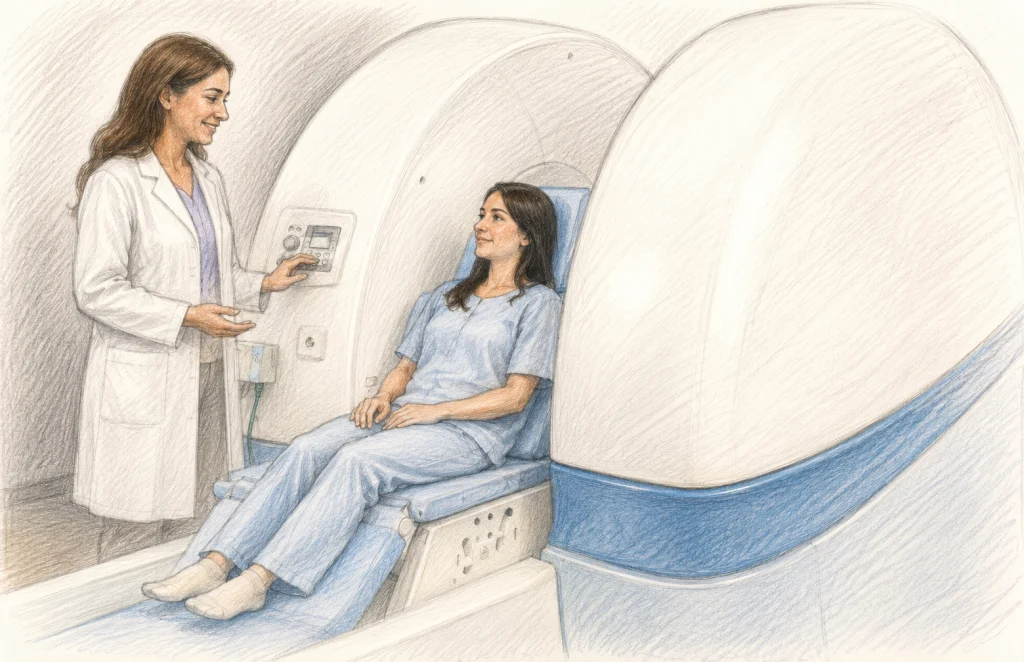
For the millions of individuals living with Ehlers-Danlos syndrome (EDS), the journey to a diagnosis is often paved with clinical frustration. A typical medical record for an EDS patient might contain a series of "unremarkable" MRI reports: a cervical spine that appears normal, a thoracic spine showing no issues, and perhaps a minor disc bulge at L5-S1 that fails to account for the patient’s debilitating, systemic neurological pain.
This disconnect between subjective patient suffering and objective diagnostic findings is a hallmark of the EDS experience. But as we move further into the era of personalized medicine, researchers and clinicians are beginning to question whether the fault lies in the patient’s biology or in the diagnostic technology itself. Specifically, is the gold standard of supine (lying down) MRI failing to capture the mechanical realities of a spine affected by connective tissue fragility?
The Mechanics of the Problem: Why Supine Imaging Often Fails
Standard Magnetic Resonance Imaging (MRI) is a marvel of modern medicine, providing unparalleled soft-tissue contrast. However, it is fundamentally a static, gravity-neutral assessment. In a standard MRI suite, the patient lies prone or supine, suspended in a state of artificial rest.
For the general population, this is sufficient. But for those with EDS—a group characterized by hypermobility, ligamentous laxity, and structural instability—the spine is often a dynamic, mechanical problem rather than a static one. In EDS, the tissues that are meant to tether and stabilize the vertebral column are inherently "stretchy." Consequently, a spine that appears aligned and stable while lying down may undergo significant shifting, translation, or collapse under the weight-bearing demands of gravity, movement, and daily upright activity.
Imaging the spine in its least symptomatic state (horizontal and unloaded) and attempting to diagnose a condition that manifests during vertical, loaded activity creates a classic "diagnostic mismatch." The technology is not flawed, but its application in the context of hypermobility may be fundamentally incomplete.
Chronology of a Shift in Diagnostic Thinking
The conversation surrounding upright MRI (often called positional or weight-bearing MRI) has evolved significantly over the last two decades.
- Early 2000s: Initial case studies began to emerge, suggesting that patients with occult spinal instability or Chiari malformations often showed anatomical shifts only when placed in an upright, weight-bearing position.
- 2007: A landmark study by Milhorat et al. highlighted the syndrome of occipitoatlantoaxial hypermobility, noting that "cranial settling" and other craniocervical junction (CCJ) abnormalities were frequently missed on standard supine scans.
- 2015–2018: Health Quality Ontario and subsequent researchers began formalizing the "state of the art" for dynamic imaging, establishing that positional MRI could reveal altered CSF flow dynamics and spinal canal narrowing that remained hidden at rest.
- 2025–2026: A wave of systematic reviews, including the 2025 scoping review on cervical spine utility, has provided a more robust evidentiary base, pushing the medical community to acknowledge that "weight-bearing" is a critical variable in assessing structural integrity in connective tissue disorders.
Supporting Data: What the Research Says
The core argument for upright MRI lies in its ability to capture "posture-dependent" pathology. While large-scale, randomized controlled trials comparing upright to supine imaging in EDS are still forthcoming, the existing literature provides compelling evidence for its utility.
A 2025 systematic review identified nine distinct studies confirming that dynamic and upright MRI can detect abnormalities—such as spinal cord compression, nerve root impingement, and altered alignment—that are invisible in supine scans. The research suggests that for patients who experience symptom relief when lying down but immediate exacerbation upon standing or head movement, the upright MRI serves as a functional stress test.
The Trade-off: Field Strength vs. Field of View
Critics of upright MRI often point to the "field strength" argument. Standard hospital-grade MRI scanners typically operate at 1.5T or 3.0T (Tesla), offering high-resolution images. Upright or open MRI systems generally operate at lower field strengths, typically 0.6T to 1.0T.
This presents a clinical trade-off:
- High-Field Supine MRI: Offers superior anatomical detail of soft tissues but fails to capture the mechanical, dynamic instability caused by gravity.
- Low-Field Upright MRI: Offers slightly lower image resolution but provides the essential "functional" data of how the spine behaves under load.
For the clinician, the diagnostic value is shifting. Many now argue that seeing the movement of the vertebrae—the way the spine "buckles" under weight—is more clinically relevant than seeing the fine texture of a disc that appears perfectly healthy in a horizontal position.
Implications for the Neurosurgical Landscape
The integration of upright MRI into the diagnostic pathway for EDS has profound implications for patient care. It changes the surgical decision-making process. If a surgeon can visualize a patient’s spinal cord being compressed specifically in an upright, flexed, or extended position, it provides a "smoking gun" for symptoms that were previously labeled as idiopathic or psychosomatic.
Organizations such as the CCI Foundation (Craniocervical Instability Foundation) have begun advocating for the inclusion of upright imaging in the evaluation of patients with suspected craniocervical instability. This is not to suggest that every patient with EDS needs an upright scan, but rather that for those whose clinical picture is discordant with their imaging, this tool provides a necessary "next step."
Access and Practical Hurdles
Despite the promise of this technology, the path to obtaining an upright MRI is often difficult. These machines are not standard equipment in most hospitals. They are often found in specialized private imaging centers, which creates two primary barriers:
- Geographical Access: Patients may need to travel across states or countries to reach a facility with upright, weight-bearing capabilities.
- Insurance and Cost: Because upright MRI is sometimes viewed as an "adjunct" or "experimental" tool rather than a standard diagnostic necessity, insurance coverage is highly variable. Many patients face the burden of out-of-pocket costs, creating a socioeconomic divide in who can access the diagnostic clarity that positional imaging offers.
When Should You Discuss Upright MRI with Your Provider?
An upright MRI may be a relevant consideration if your clinical symptoms do not match your current imaging results. Specifically, consider this pathway if:
- Your symptoms (headaches, nerve pain, dizziness, limb weakness) correlate strictly with standing or sitting.
- You experience "head heaviness" or mechanical neck pain that worsens throughout the day.
- You have a confirmed diagnosis of an EDS subtype and are experiencing neurological decline that standard imaging cannot explain.
- You have been told your cervical spine is "normal," yet you struggle with autonomic symptoms or balance issues that suggest brainstem or spinal cord irritation.
Key Takeaways for Patients and Providers
- The Spine is Dynamic: It is not a static bridge; it is a weight-bearing structure that reacts to gravity. In EDS, the "normal" load of daily life can induce pathological movement.
- A Complement, Not a Replacement: Upright MRI should not replace standard high-field MRI. Instead, it should be viewed as a functional diagnostic aid that provides a different, complementary perspective.
- Advocacy is Essential: Because the technology is not yet universally adopted, patients must be proactive in discussing the limitations of supine imaging with their specialists.
- Future Research: While current data is promising, the field needs larger, multi-center studies to standardize the criteria for "instability" on upright scans to ensure that these results are interpreted accurately by radiologists and neurosurgeons alike.
Conclusion
The medical community is slowly recognizing that in the world of connective tissue disorders, our current diagnostic window is too narrow. By relying exclusively on horizontal imaging, we have been asking patients to demonstrate their pathology in a state where their body is most supported.
As we look to the future, the goal is to bridge the gap between patient experience and clinical observation. Upright MRI represents a significant step forward, offering a more nuanced view of how the spine navigates the daily forces of gravity. By improving access to this technology and continuing to fund research into its clinical utility, we can ensure that patients with EDS are no longer told their imaging is "unremarkable," but rather, that they are finally being seen in a way that captures the reality of their condition.
When Might Upright MRI Be Relevant for You?
- Postural Dependency: When symptoms (e.g., vertigo, radiculopathy, or severe neck pain) consistently resolve when you lie down.
- Discordant Findings: When you have severe, documented neurological symptoms but your previous MRI scans were reported as "normal."
- Suspected CCI: If you are being evaluated for craniocervical instability or Chiari malformation and standard scans are inconclusive.
- Mechanical Instability: If you feel "clicking," "grinding," or a sense of "giving way" in your spine when changing positions.
Key Takeaways
- Gravity Matters: Supine MRI ignores the weight-bearing stress that often triggers symptoms in EDS patients.
- Dynamic Imaging: Upright MRI acts as a functional test, capturing the spine’s movement under gravity.
- The Trade-off: Lower image resolution on some upright systems is often an acceptable trade for the high-value clinical data gained regarding spinal stability.
- Patient Advocacy: Due to limited access and insurance hurdles, patients often need to actively coordinate with their healthcare team to justify the necessity of positional imaging.