










By John Gever, Contributing Writer, MedPage Today
October 15, 2026
In the management of systemic sclerosis (SSc)—a complex autoimmune condition characterized by the hardening and tightening of the skin and connective tissues—clinicians have long relied on the "modified Rodnan Skin Score" (mRSS). This subjective physical examination involves a physician manually pinching and assessing skin thickness across various body sites. While it remains the clinical gold standard, it is prone to inter-observer variability and lacks the granular sensitivity required to detect subtle disease progression or therapeutic response.
However, a breakthrough in noninvasive, nonradiological imaging may soon change this. A new study published in The Lancet Rheumatology highlights the potential of spatial frequency domain imaging (SFDI) as a powerful, objective tool for evaluating skin fibrosis. Led by Dr. Andreea M. Bujor of Boston University (BU), the research suggests that SFDI could eventually replace or supplement the Rodnan score, offering a more precise way to track the trajectory of scleroderma over time.
The Limitations of Current Assessment
To understand the significance of this development, one must appreciate the diagnostic hurdles inherent in SSc. Because fibrosis in systemic sclerosis occurs deep within the dermis and subcutaneous layers, visual inspection is insufficient. The current gold standard, the mRSS, is a manual, "hands-on" assessment that is inherently subjective.
When physicians require objective proof of disease activity, they are forced to rely on histological biopsies—a process that is invasive, painful for the patient, and physically impossible to perform with the frequency needed to capture real-time disease fluctuations. As a result, many patients go months or even years without an accurate assessment of whether their disease is worsening or responding to treatment, leaving clinicians to make decisions based on delayed or incomplete snapshots of the patient’s condition.
What is Spatial Frequency Domain Imaging (SFDI)?
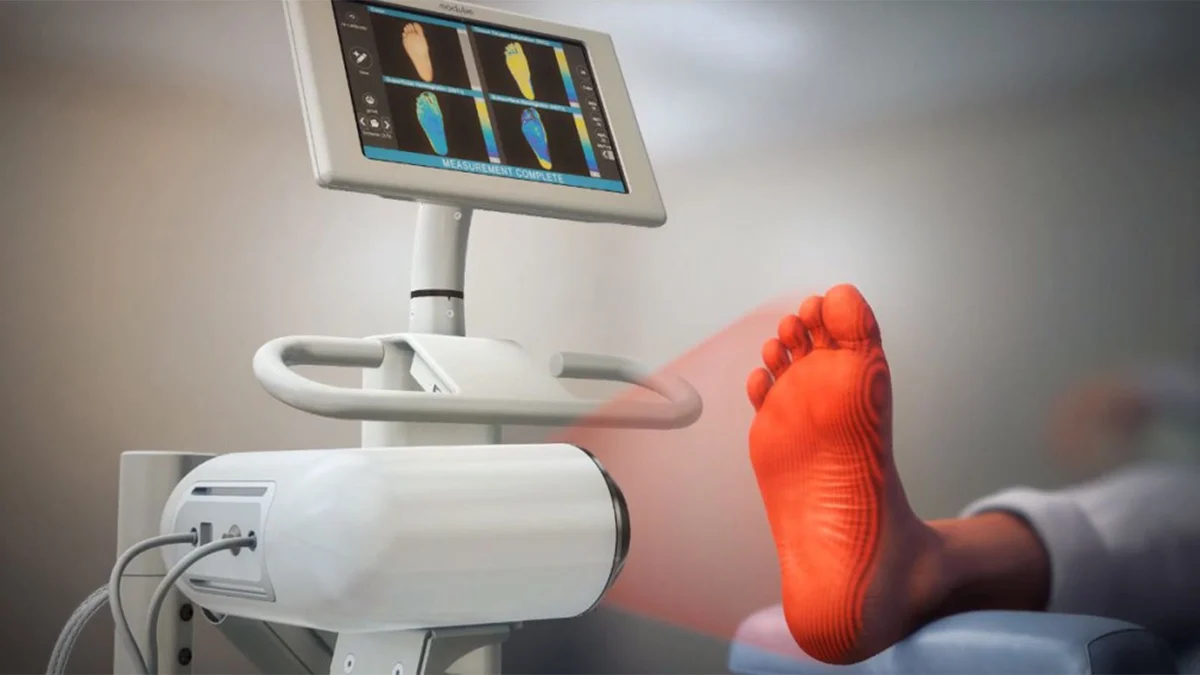
First introduced in 2009, SFDI was designed as a method for the wide-field, quantitative characterization of the optical absorption and scattering properties of turbid media—biological tissues that scatter light. Unlike X-rays or CT scans, SFDI uses ordinary, safe visible light to map the physical properties of the skin.
By projecting structured light patterns onto the skin and analyzing how the tissue reflects and scatters that light, SFDI provides a high-resolution map of tissue health. It penetrates the layers of the skin to measure structural changes associated with fibrosis, providing data that can be translated into numerical values. Because the procedure is noninvasive and radiation-free, it allows for the kind of frequent serial monitoring that biopsy-based assessment simply cannot facilitate.
Chronology of the BU Clinical Trial
The study conducted by Dr. Bujor and her colleagues was designed to determine whether these optical measurements correlate with the traditional clinical markers of SSc.
Phase 1: The Pilot Study
The investigation began with a pilot phase involving 10 patients with SSc and eight healthy controls. These participants underwent comprehensive testing, including ultrasound, durometry, and SFDI scans during a single visit. To validate the findings, the researchers offered participants the option of forearm biopsies, providing a biological baseline to compare against the optical data.
Phase 2: The Longitudinal Expansion
Building on the promising results of the pilot, the team expanded the cohort. The final analysis included 49 SSc patients and 25 healthy controls. In this phase, the researchers moved beyond static snapshots to longitudinal observation. Participants were monitored every three months for the first year, and every six months for an additional three years. This multi-year window allowed the team to assess whether SFDI could detect "within-patient change" over time—the holy grail of chronic disease monitoring.
Supporting Data: The Correlation Strength
The results of the study were striking. The researchers tracked two primary SFDI parameters: diffuse reflectance ($R_d$) and the reduced scattering coefficient ($mu_s’$).
When compared to the mRSS, these parameters demonstrated a robust correlation (with $r$ coefficients ranging from -0.38 to -0.48). Even more compellingly, the correlation between SFDI values and histological findings of fibrosis—the actual physical scarring of the tissue under a microscope—showed a correlation range of -0.34 to -0.52.
Perhaps most importantly, the researchers observed that values for both $R_d$ and $mu_s’$ were significantly lower in patients with scleroderma compared to healthy controls, regardless of their current Rodnan score. This suggests that SFDI can identify pathological tissue changes that the human touch—even that of an expert clinician—might miss.
Expert Insights and Official Responses
Dr. Bujor and her team are careful to temper their excitement with scientific rigor. While the study results are promising, they acknowledge that the sample size, while adequate for a preliminary study, is not yet representative of the full diversity of the SSc population.
"Changes in SFDI parameters correlated with changes in mRSS, suggesting that SFDI can track within-patient change over time," the authors noted in their report. "However, because the longitudinal sample was modest and change-score analyses can be influenced by regression to the mean, these findings should be interpreted cautiously and confirmed in larger prospective cohorts."
The researchers highlighted that the current study did not account for several potential confounders, such as variations in medication use, smoking status, or sun exposure, all of which could influence skin optical properties. Nevertheless, the ability to collect longitudinal data over four years provided a level of detail that has previously been unavailable in SSc clinical research.
Future Implications: The Path to Clinical Adoption
For SFDI to move from a research laboratory to a clinical setting, several hurdles must be cleared. The researchers identified four key priorities for future development:
- Handheld Optimization: The current equipment must be refined into a portable, user-friendly device that can be easily operated in a standard rheumatology clinic.
- Validation in Diverse Cohorts: The technology must be tested across a broader range of patients, spanning from those with "very early" disease to those with long-standing, chronic skin involvement.
- Thresholds of Meaning: Clinicians need to know exactly what a specific change in an SFDI score means for the patient. Establishing "minimal clinically important differences" (MCID) will be vital.
- Patient-Reported Outcomes: The optical data must be reconciled with how patients actually feel and function. Integrating SFDI results with patient-reported quality-of-life metrics will be the final step in proving its clinical utility.
If successful, the implications for systemic sclerosis are profound. Beyond SSc, researchers believe this technology could be adapted for a wide variety of other skin diseases where fibrosis or inflammation plays a role, including morphea or systemic lupus erythematosus.
By shifting the paradigm from the "pinch" to the "pixel," the medical community moves one step closer to a future where skin health is measured with the same precision as blood pressure or blood glucose—transforming the management of a once-mysterious disease into a data-driven, highly responsive standard of care.
For now, the rheumatology community will watch closely as Dr. Bujor’s team and others work to validate these findings. If the promise holds, the subjective era of the Rodnan score may eventually give way to an age of objective, optical, and noninvasive precision medicine.