


For decades, patients living with hypermobile Ehlers-Danlos syndrome (hEDS) have faced a demoralizing medical paradox. They report debilitating symptoms—chronic migraines, persistent nausea, vertigo, "brain fog," severe neck pain, and profound fatigue—that often leave them bedbound. Yet, when these patients seek answers through imaging, they are frequently told their results are "normal."
For years, the medical community lacked a standardized definition of "normal" regarding the craniocervical junction (the area where the skull meets the spine). Diagnosis often relied on the subjective, inconsistent observations of individual specialists, leaving patients trapped in a diagnostic limbo.
However, the landscape of hEDS care is beginning to shift. Two landmark studies published in 2025 have provided the first rigorous, data-driven frameworks to analyze the relationship between hEDS and the upper cervical spine. These findings do not just offer academic data; they represent a significant step toward moving hEDS diagnosis away from subjective guesswork and toward objective, standardized clinical practice.
The Core Problem: Why "Normal" Has Been a Moving Target
The frustration experienced by hEDS patients is rooted in the absence of a universal "gold standard" for imaging the craniocervical junction. Because connective tissue disorders like hEDS affect the body’s structural integrity, the ligaments that hold the skull and the first two vertebrae (C1 and C2) in place may be lax. This can lead to craniocervical instability (CCI) or atlanto-axial instability (AAI).
Historically, radiologists and neurosurgeons have relied on internal, non-standardized measurements to assess whether a patient’s neck was "stable." A measurement deemed "normal" by one physician might be viewed as pathological by another. Patients suffering from severe neurological symptoms were routinely dismissed because their scans fell within the "normal" range of a specific clinic’s internal secret code. This lack of consensus has left many patients feeling gaslit, their pain minimized, and their structural concerns ignored.
Chronology of Progress: A Shift in Diagnostic Standards
The path to these recent breakthroughs began with the urgent need for a shared clinical language.
- Early 2000s–2020: The medical community struggled with high variability in imaging results. Experts often debated whether symptoms were truly structural or merely functional.
- 2024: Researchers began finalizing studies aimed at quantifying the "normal" cervical spine using diverse imaging modalities.
- 2025: Two pivotal papers were published, addressing the critical questions of baseline normalcy and the prevalence of structural variations in the hEDS population.
- 2026 and Beyond: These studies provide the foundational data necessary to train AI-driven diagnostics and update medical guidelines, potentially reducing the time to diagnosis for thousands of patients.
Supporting Data: Defining the Boundaries of Normalcy
The first study, titled Radiographic Indicators of Craniocervical Instability: Analyzing Variance of Normative Supine and Upright Imaging in a Healthy Population, sought to define the baseline. Researchers analyzed seven critical anatomical markers in 72 healthy individuals.
By measuring these markers across four different positions—extension x-ray, flexion x-ray, neutral x-ray, and supine CT scans—the researchers discovered that "normal" is highly dependent on how the image is captured.
The Seven Key Metrics
The study examined:
- Clivo-axial angle (CXA): The alignment of the skull base and the spine.
- Basion-dens interval (BDI): The gap between the skull base and the C2 vertebra.
- Basion-axis interval (BAI): The front-to-back alignment of the skull base relative to the spine.
- Atlanto-dental interval (ADI): The critical space between the C1 and C2 vertebrae.
- Perpendicular basion to the inferior aspect of C2 (pbC2): Often called the Grabb-Oak measurement, this tracks how far the skull base projects toward the spinal canal.
- Hard palate to C1 (HPC1): The relationship between the nasal cavity and the C1 vertebra.
- Hard palate to C2 (HPC2): The relationship between the nasal cavity and the C2 vertebra.
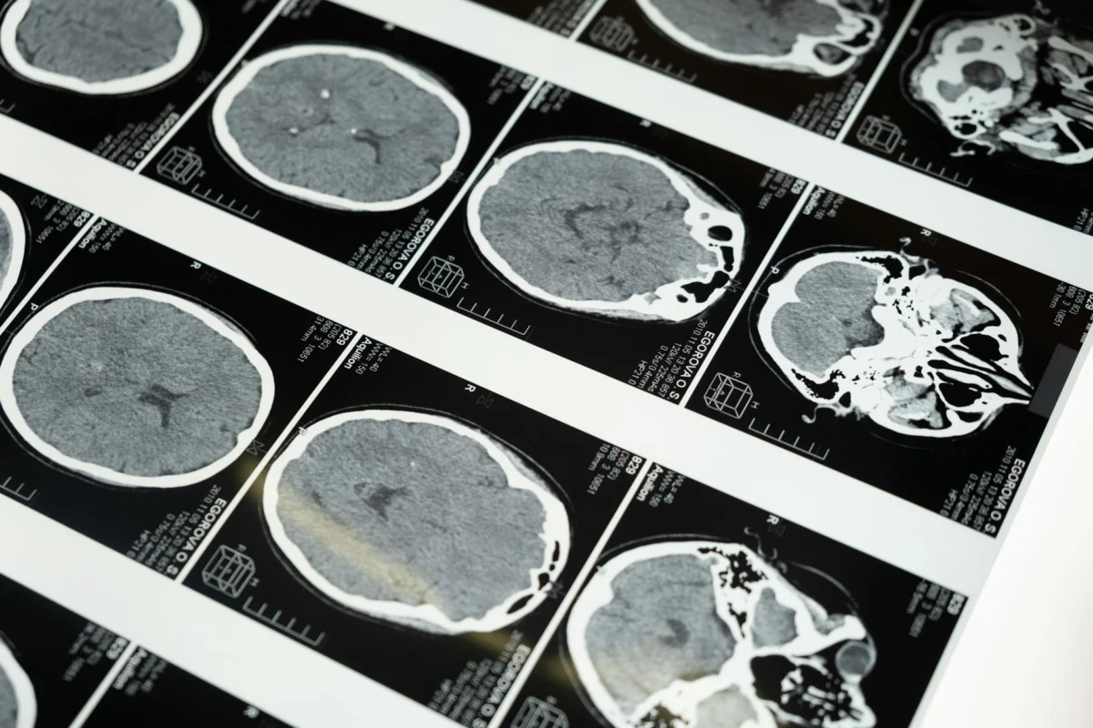
The study revealed that the CXA (Clivo-axial angle) is the most reliable metric across all imaging types. This discovery provides a more consistent "anchor" for clinicians. Crucially, the researchers noted that a measurement that appears normal on a CT scan might deviate significantly on an extension x-ray. This underscores the importance of functional imaging—taking pictures while the patient is moving or in different positions—rather than relying solely on static, supine imaging.
Structural vs. Postural: The hEDS Difference
The second major study, Head Posture and Upper Spine Morphological Deviations in People With Hypermobile Ehlers-Danlos Syndrome, tackled the question of whether hEDS patients have inherently different anatomy.
Using lateral cephalograms and cone-based computed tomography (CBCT), researchers compared 27 individuals with hEDS against 39 individuals without. The results were surprising: there were no statistically significant differences in basic head posture or anatomical measurements between the two groups. This suggests that the symptoms experienced by hEDS patients are not necessarily the result of a "different" starting structure, but rather how that structure behaves under stress.
The Significance of Anatomical Deviations
While the baseline structure was similar, the prevalence of deviations was not. Over half (51.9%) of the hEDS cohort displayed anatomical variations, compared to only 15% of the control group.
Most notably, researchers identified a high frequency of posterior arch deficiencies (PAD)—developmental gaps in the posterior arch of the C1 (atlas) vertebra. While these are often categorized as "spina bifida occulta" and dismissed as asymptomatic, their disproportionate prevalence in the hEDS group suggests they may be a structural marker that, when combined with ligamentous laxity, predisposes a patient to symptomatic instability.
Implications for Clinical Practice
These findings serve as a wake-up call for the medical community. The implications for patient care are profound:
- Standardization of Imaging: Clinicians can no longer rely on a single, static CT scan to rule out structural issues. The clear differences between supine, neutral, and flexion/extension imaging mean that protocols must be updated to include functional views when a patient presents with neurological symptoms.
- Refined Diagnostic Criteria: The identification of posterior arch deficiencies as a potential marker for hEDS indicates that we are moving toward a more objective, phenotype-based diagnostic model. While genetic markers for hEDS remain elusive, physical markers in the upper cervical spine can now be used as "clinical flags."
- Validation of Patient Experience: Perhaps most importantly, these studies provide a scientific basis for the symptoms patients have described for years. By proving that structural variations are significantly more common in hEDS patients, the research effectively ends the debate over whether these symptoms are "all in their heads." While the symptoms are in the head and neck, they are now confirmed to be rooted in quantifiable, measurable physical deviations.
Moving Forward: A Foundation, Not a Finish Line
These studies represent a watershed moment in the management of Ehlers-Danlos syndrome. By defining the parameters of the healthy craniocervical junction and identifying the anatomical patterns associated with hEDS, researchers have provided the tools necessary to move beyond the era of dismissal.
However, it is vital to remember that these results are a starting point. Future research must expand the diversity of study populations—ensuring that transgender, non-binary, and a wider range of ethnic groups are included—to ensure these "normal" ranges are truly universal.
For the patient who has spent years being told their pain was "normal," these papers offer more than just data. They offer vindication. They signal that the medical community is finally looking in the right place, with the right tools, and with a growing commitment to seeing the person behind the symptoms. The path to better care for hEDS is no longer a mystery; it is a clear, evidence-based road that is finally under construction.