










Every year, seasonal influenza imposes a staggering burden on global public health, accounting for 3 to 5 million cases of severe respiratory illness and up to 650,000 deaths. Beyond the predictable annual cycle, the Influenza A virus remains a perennial candidate for global pandemics, a legacy punctuated by the devastating 1918 Spanish Flu. Despite decades of study, the precise molecular "handshake" between the virus and its host has remained elusive—until now.
In a landmark study published in Nature Microbiology, researchers from EMBL Hamburg and the Leibniz Research Institute for Molecular Pharmacology (FMP) have unveiled an unprecedented, high-resolution map of how Influenza A systematically deconstructs and hijacks human cellular machinery. By observing protein interactions within intact, living cells, the team has moved beyond the "broken-cell" limitations that have hampered virology for decades, opening a new frontier in the development of next-generation antiviral therapies.
The Challenge of the "Broken" View
To understand why this discovery is revolutionary, one must understand the historical constraints of molecular biology. For years, the gold standard for mapping protein-protein interactions (PPIs) involved biochemical techniques that required researchers to lyse—or break open—infected cells.
While effective at identifying which proteins were present in a sample, the method was inherently flawed. By destroying the structural integrity of the cell, researchers inadvertently removed the biological context of the interactions. Proteins that were naturally sequestered in different organelles were suddenly thrown together in a test tube, creating "false positive" interactions. Conversely, weak, transient, or location-specific interactions—the very mechanisms the virus uses to replicate—were often lost in the chaos of the lysis process.
"The current results are a snapshot of a moment during infection, and it opens the door to studying flu-host interactions across the entire infection cycle," explains Jan Kosinski, Group Leader at EMBL Hamburg and the Centre for Structural Systems Biology (CSSB). By capturing the virus in its native habitat, the researchers have finally bridged the gap between theoretical models and the messy reality of an active infection.
Chronology: A Multi-Institutional Breakthrough
The success of this project was not a solitary endeavor but a masterclass in interdisciplinary collaboration across three major German research hubs.
Phase I: Adapting the Toolset
The foundation was laid when the EMBL team partnered with Boris Bogdanow and Fan Liu at FMP Berlin. The researchers utilized a specialized evolution of cross-linking mass spectrometry (XL-MS). Unlike traditional mass spectrometry, XL-MS uses chemical "staples" to lock proteins together while they are still inside the living cell. This preserves the exact spatial relationship between viral and host proteins before the cell is processed.
Phase II: The Computational Integration
Once the cross-linked data was captured, the team faced a massive structural puzzle: how do these proteins physically fit together? To solve this, they integrated their experimental data with a modified version of AlphaFold, the Nobel Prize-winning protein structure prediction algorithm. By feeding the cross-linking "constraints" directly into the algorithm, the researchers were able to model the virus-host complexes with high confidence, identifying specific interfaces that were previously invisible.
Phase III: Validation and Imaging
The team utilized the EMBL Compute Cluster to process the structural models, while researchers at the CSSB’s Advanced Light and Fluorescence Microscopy (ALFM) Facility provided visual validation. This final layer of microscopy confirmed that the structural predictions matched the biological reality occurring within the nucleus and the cytoplasm of the infected cells.
Decoding the Hijacking Strategies
The study identified two primary, distinct strategies that Influenza A employs to dismantle cellular defenses.
The Hemagglutinin Processing Network
Influenza A relies on a surface protein called hemagglutinin to infiltrate the host cell. Once inside, the virus must fold and modify this protein to ensure the new viral particles are functional. The study revealed that the virus does not act alone; it actively recruits host cell proteins to assist in the "quality control" of hemagglutinin.
These human proteins, which typically handle protein folding in the cell’s internal transport network, are repurposed by the virus. By mapping these interactions, the scientists identified several human factors whose roles in infection were previously unknown, providing potential "choke points" where drugs could theoretically block the virus from finalizing its own production.
The Dissolution of Paraspeckles
Perhaps the most startling discovery involved "paraspeckles"—tiny, droplet-like compartments within the cell nucleus. The team observed that the Influenza A virus consistently causes these structures to dissolve.
"Watching these tiny organelles in the nucleus dissolve, consistently across every cell line and every flu strain we tested, told us this isn’t a side effect of infection—it might be a strategy," says Iuliia Kotova, the study’s first author and a former predoctoral fellow at EMBL Hamburg.
The dissolution of paraspeckles appears to serve a dual purpose. First, it releases RNA-binding proteins that the virus then hijacks to support its own replication. Second, because paraspeckles are vital to the cell’s stress response and antiviral gene regulation, their destruction effectively blinds the cell, preventing it from mounting a coordinated immune defense.
Supporting Data and Technical Precision
The rigor of the study is supported by the sheer volume of data integrated into the final model. The team’s approach involved:
- XL-MS Capture: Identifying thousands of physical contact points between viral and host proteins.
- Glycoproteomics: Performed at the EMBL Proteomics Core Facility, this analysis allowed the researchers to understand the chemical modifications on the proteins that influence their interaction.
- AlphaFold Modeling: The integration of experimental data allowed for the structural modeling of virus-host interfaces, a feat that is notoriously difficult for standard computational models which lack in-cell spatial constraints.
Official Responses and Scientific Impact
The research community has noted the work as a paradigm shift. By moving from a "list" of proteins to a "map" of interactions in space and time, the authors have provided a blueprint for how to study other viral threats.
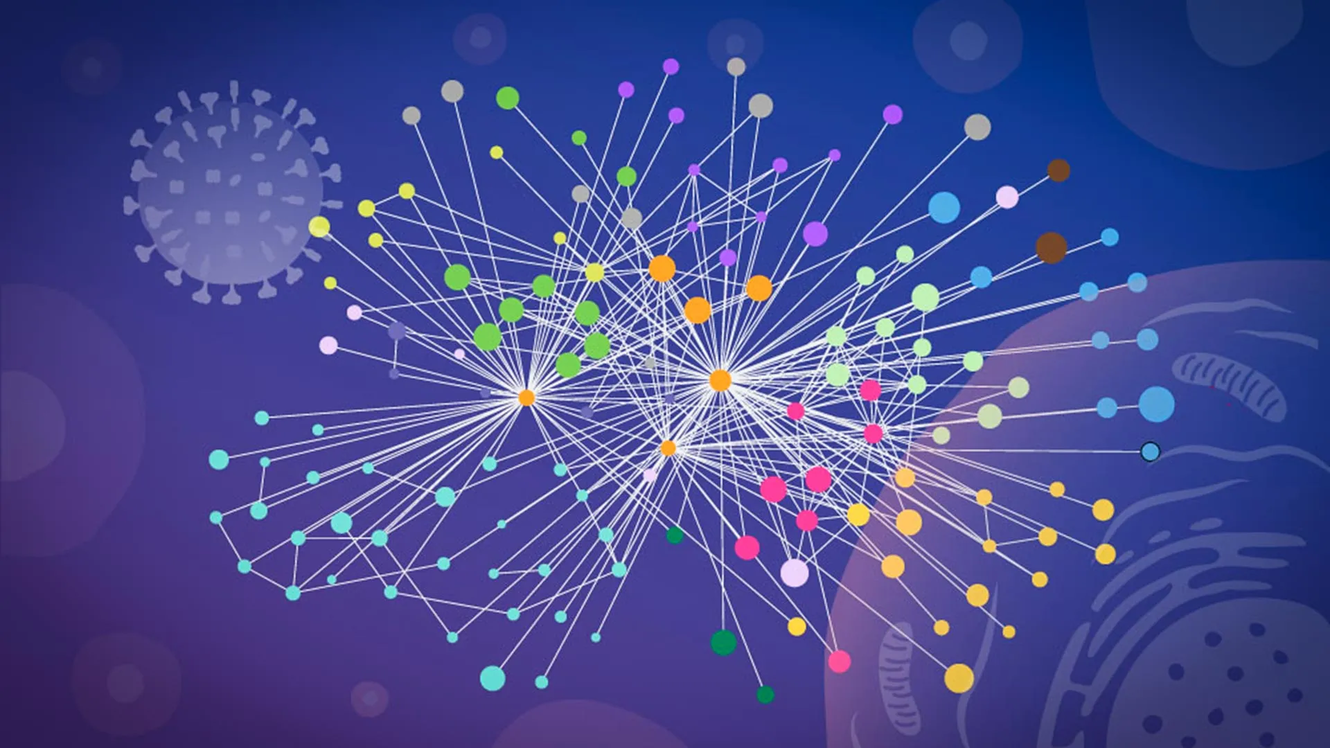
"This gives us insight into the interface between the virus and the human cell and may, through structural modeling, help identify actionable targets for future pharmaceutical interventions," said Boris Bogdanow, now a Junior Research Group Leader at the Institute of Virology at Charité – Universitätsmedizin Berlin.
The implications for drug development are significant. Current antivirals often target the virus directly, leading to rapid mutations and drug resistance. By identifying the human proteins that the virus requires for survival, researchers may be able to develop "host-targeted" therapies. Because these human proteins are less prone to mutation than viral proteins, this strategy could create a higher barrier to resistance.
Future Implications: From Lab Strains to Pandemics
While this study focused on a laboratory-adapted strain of Influenza A, the researchers are already looking toward the horizon. The methodology—combining in-cell cross-linking, structural modeling, and targeted cell-biology follow-up—is inherently scalable.
The team has expressed a strong desire to apply these techniques to high-pathogenicity viruses, such as H5N1. As these viruses continue to circulate in avian and mammalian populations, the ability to rapidly map their interaction networks could be the difference between a controlled outbreak and a global pandemic.
"While the exact host factors and mechanisms often differ from virus to virus, we think our overall approach… remains broadly applicable," says Kosinski.
By illuminating the "dark matter" of virus-host interactions, this research has provided the scientific community with a new set of tools to see not just what the virus is, but how it thinks, how it moves, and ultimately, how it can be stopped. As the world remains vigilant against the next potential pandemic, the work at EMBL and FMP stands as a testament to the power of integrating structural biology with live-cell imaging to solve one of the most complex puzzles in medicine.