









By [Your Name/Editorial Staff]
For those living with narcolepsy type 1, the most profound moments of human connection—a sudden burst of laughter at a dinner table, the thrill of a surprise visit from an old friend, or the joy of a competitive game—are frequently shadowed by a sudden, involuntary loss of muscle tone known as cataplexy. For decades, patients have reported this paradoxical phenomenon: their happiest moments are precisely when their bodies are most likely to fail them.
While this pattern has been clinically recognized for years, the underlying physiological "why" remained elusive. Now, a groundbreaking study led by researchers at Harvard Medical School has finally bridged the gap between emotional reward and physical paralysis, pointing to a specific neural circuit governed by oxytocin—the very hormone responsible for love, bonding, and social trust.
The Intersection of Emotion and Physiology: Main Facts
At its core, narcolepsy type 1 is a chronic neurological condition characterized by the brain’s inability to regulate sleep-wake cycles. A defining hallmark of the disorder is cataplexy, a sudden muscle weakness triggered by strong emotions.
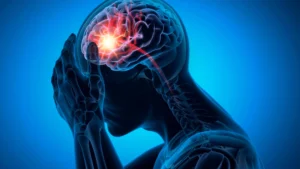
The new research, published recently, identifies oxytocin as the critical link in this chain. By utilizing a mouse model of narcolepsy, the team demonstrated that when an individual experiences a positive social interaction, the brain releases oxytocin. In the narcoleptic brain, this surge of oxytocin does not merely facilitate bonding; it activates a specific pathway that inadvertently suppresses the brainstem mechanisms responsible for maintaining muscle tone. Essentially, the brain’s "social reward" system hijacks the body’s "muscle control" system.
A Chronological Breakdown of the Discovery
The research process was a multi-stage endeavor that combined behavioral observation with sophisticated optogenetic mapping.
Phase 1: Isolation and Reunification
The team first sought to replicate the human experience of socially triggered cataplexy in a controlled environment. They implemented a social isolation and reunification test. Mice were separated from their cage mates for a brief period. When the animals were reunited, the researchers observed a statistically significant spike in cataplectic episodes compared to baseline measurements. By carefully adjusting variables, the team ruled out confounding factors such as the stress of handling or the novelty of the environment, confirming that the social interaction itself was the primary catalyst.
Phase 2: Testing the Oxytocin Hypothesis
To confirm the role of oxytocin, the researchers employed a two-pronged pharmacological approach. First, they introduced an oxytocin receptor antagonist—essentially a chemical "blocker"—to the mice. Upon doing so, the socially triggered cataplexy was significantly averted. Conversely, when they administered an oxytocin receptor agonist (carbetocin), which mimics the hormone’s effects, the frequency and duration of cataplexy bouts increased dramatically compared to a saline-controlled group.
Phase 3: Mapping the Circuitry
With the hormone identified, the next hurdle was locating the physical path. Using fiber photometry and neural sensors, the team mapped the projections from the paraventricular nucleus of the hypothalamus to the central amygdala—a region already known for its role in regulating muscle tone. They discovered that oxytocin-sensitive neurons in this pathway effectively "shut down" the protective neurons in the brainstem that maintain muscle atonia, thereby triggering the physical collapse.
Supporting Data: The "Reward" Overlap
One of the most intriguing aspects of the study was the investigation into whether this circuit is exclusive to social bonding or if it extends to broader reward-based experiences. To test this, researchers offered the mice milk chocolate.
The results were telling: the chocolate triggered the same oxytocin-amygdala pathway, inducing cataplexy. This suggests that the brain does not distinguish between the "reward" of a social interaction and the "reward" of a high-value treat. Both stimuli recruit the same neuronal machinery. Furthermore, using real-time place preference tests, the researchers confirmed that the mice found the stimulation of these neurons inherently rewarding, reinforcing the link between the brain’s pleasure centers and the onset of cataplexy.
Official Perspectives and Expert Insight
Dr. Carrie Mahoney, an instructor in neurology in the Division of Sleep Medicine at Harvard Medical School and the study’s corresponding author, emphasizes that this research is not merely about finding a new biological marker—it is about validating the lived experience of patients.
"We had a feeling it was either oxytocin or some other social-related hormone that was going to increase cataplexy, but we weren’t sure," Dr. Mahoney explains. "We drilled even further down to a subpopulation of neurons, trying to understand the oxytocin-sensitive cells within that broader inhibitory population. We are contributing the specificity of oxytocin-sensitive neurons to the pathway."
Regarding the clinical weight of these findings, Dr. Mahoney is clear: "It’s a physical circuit, which is difficult for patients to be able to address in the moment. Their symptoms are valid, and we need to help them be able to live as normal a life as possible by helping them develop therapies to address it."
Clinical and Therapeutic Implications
The discovery of this pathway opens a new frontier in narcolepsy treatment, though it comes with significant caveats. Because oxytocin is essential for healthy social functioning, simply blocking it in humans is not a viable therapeutic strategy. As Dr. Mahoney notes, "You don’t want to interrupt normal human behavior or normal social interactions… You could potentially limit the benefits of social interaction, which we would not want to do at all."
Instead, the research points toward a more granular approach:
- Targeted Molecular Intervention: By identifying the specific biological markers of these oxytocin-sensitive neurons, researchers hope to develop drugs that inhibit only the "cataplexy-triggering" sub-population without interfering with the rest of the hormone’s social benefits.
- Imaging and Diagnostics: The identification of this pathway may allow clinicians to use fMRI and other neuroimaging tools to better characterize the severity of a patient’s condition.
- Addressing Sex Differences: The study unexpectedly revealed that juvenile female mice experienced twice as many cataplectic episodes as their male counterparts. This observation suggests that developmental hormonal fluctuations could influence symptom severity, a topic that has received insufficient attention in human clinical trials.
Looking Ahead: The Path Toward New Treatments
The journey from a mouse model to a clinical treatment for humans is long and fraught with complexity. Unlike mice, humans possess complex cortical structures that modulate their reactions to stimuli. However, this study provides the clearest roadmap to date for why current therapies may be limited and where future breakthroughs might lie.
Dr. Mahoney is currently seeking further funding to continue exploring the molecular profile of these oxytocin-sensitive neurons. Her goal is to identify an alternative therapeutic target—a "switch" that, if toggled, could allow patients to enjoy a laugh with friends or a moment of triumph without the fear of an sudden, involuntary collapse.
For the narcolepsy community, this research represents more than just a scientific breakthrough; it represents a fundamental validation of their reality. By proving that cataplexy is a hard-wired physiological response to the very things that make life worth living, science has taken a massive step toward de-stigmatizing the disorder and providing a future where patients can experience joy without restriction.