







For over a century, a quiet anatomical phenomenon has drifted in and out of the medical consciousness. Known as "visceroptosis"—the downward displacement of abdominal organs—it is a condition that once occupied the pages of the world’s most prestigious medical journals, only to fade into relative obscurity, often dismissed as a byproduct of "nervous" temperament. Today, however, as our understanding of connective tissue disorders like Hypermobile Ehlers-Danlos syndrome (hEDS) deepens, the medical community is finding that what was once written off as a psychological quirk may, in fact, be a significant, overlooked structural reality.
Understanding Visceroptosis: The Mechanics of Displacement
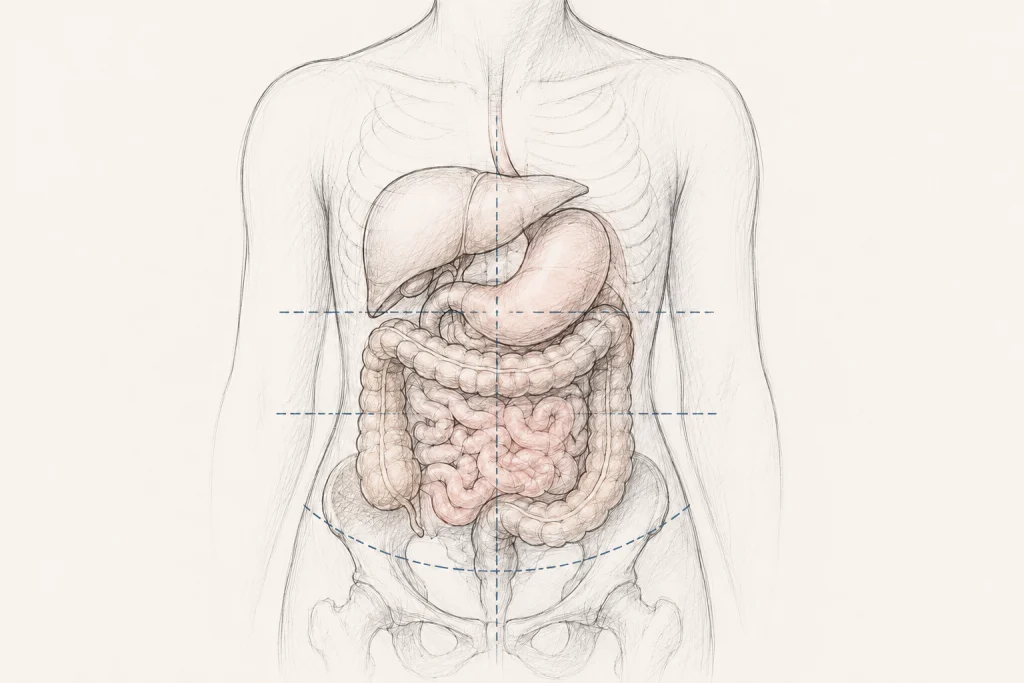
At its core, the term "visceroptosis" is derived from two Latin and Greek roots: viscera, referring to the internal organs of the abdominal and pelvic cavities, and ptosis, meaning a downward fall or displacement.
In a healthy anatomy, organs are held in a precise, suspended state by a complex network of ligaments, mesenteries, and surrounding musculature. When this support system loses its structural integrity, organs—including the stomach, liver, kidneys, and transverse colon—can shift downward, sometimes resting significantly lower in the abdominal cavity than anatomically expected. While some degree of anatomical variation is normal, visceroptosis implies a degree of descent that threatens to disrupt the harmonious function of the digestive and urological systems.
A Chronological Perspective: From "Glénard’s Disease" to Modern Neglect
The clinical history of visceroptosis is a study in medical shifting tides.
The Late 19th Century: The Rise of Diagnosis
In 1885, the French physician Dr. Frantz Glénard brought this condition to the forefront of medical literature. Glénard observed that many of his patients, particularly those complaining of chronic fatigue, abdominal pain, and digestive distress, exhibited organs that had effectively "slipped" from their moorings. For a time, the condition was eponymously known as "Glénard’s disease." During this era, physicians were diligent in their examinations; radiologists of the early 20th century actively sought signs of displacement, and surgeons were eager to explore corrective interventions. It was a standard topic of conversation in publications such as The New England Journal of Medicine, JAMA, and The Lancet.
The Mid-Century Retreat
Following the 1930s, however, the diagnosis began a steady decline. Why did a condition that appeared to cause tangible suffering suddenly fall out of clinical favor? Historians of medicine suggest a confluence of factors: the rise of psychosomatic medicine, which increasingly categorized unexplained abdominal pain as "neurasthenia" or anxiety-driven, and a lack of standardized surgical outcomes. By the mid-20th century, the medical establishment had largely moved on, viewing visceral position as a secondary, irrelevant detail compared to the burgeoning fields of pharmacology and systemic disease management.
The Contemporary Resurgence
The 21st century has seen a modest but persistent revival of interest, largely driven by the patient-advocacy community and the growing recognition of connective tissue disorders. As researchers investigate the systemic manifestations of hEDS, they have begun to find that the very fragility of collagen—the "glue" that holds the body together—naturally predisposes patients to organ displacement. What was once a "forgotten diagnosis" is now being re-examined through the lens of modern genetics and biomechanics.
Supporting Data: The Link to Connective Tissue Laxity
While the medical literature on visceroptosis in the context of EDS remains in its infancy, the data points toward a compelling correlation.
The 2017 diagnostic criteria for hEDS already acknowledge specific forms of visceral prolapse, such as rectal and pelvic organ prolapse. These conditions are now accepted as part of the phenotypic spectrum of the syndrome. However, the abdomen is a crowded space; the descent of the transverse colon or the stomach (gastroptosis) is now surfacing in case reports and smaller clinical studies.
A pivotal 2023 study published in PM&R (and widely referenced in current research) examined 139 patients with chronic, slow-transit constipation compared to 125 healthy controls. The study revealed a statistically significant association between the degree of transverse colon ptosis and the severity of symptoms. While this study did not focus exclusively on hEDS, it provided the empirical weight needed to argue that organ position is not merely an incidental finding; it is a clinical factor that influences disease severity and surgical necessity.
The Diagnostic Dilemma: Clinical vs. Anatomic Visceroptosis
A major hurdle in integrating visceroptosis into modern practice is the distinction between anatomic and clinical manifestations.
It is entirely possible for an individual to have an organ that sits lower than the "textbook" average without experiencing any symptoms whatsoever. This is anatomic visceroptosis. Conversely, clinical visceroptosis occurs when that displacement compromises physiology—impairing peristalsis, causing kinking of the bowel, or leading to debilitating pain.
Medical professionals are cautioned that the extent of descent should not be the sole diagnostic criteria. A patient’s symptoms must be carefully correlated with the anatomical findings after other common organic diseases—such as inflammatory bowel disease or malignancy—have been ruled out. This requires a shift in diagnostic imaging: standard GI evaluations are built to find tumors or inflammation, not to assess the spatial orientation of organs within the cavity.
Implications for Patients: The Struggle for Validation
The implications for patients, particularly those living with Ehlers-Danlos syndrome, are profound. For decades, many have been told that their symptoms of early satiety, bloating, and chronic pain are merely "functional" or purely psychological.
The Pattern of Dismissal
The historical description of patients with visceroptosis in the 1920s mirrors the modern experience of many hEDS patients. Early physicians labeled these individuals as "neurasthenic," a term that has evolved into modern labels like "somatic" or "anxious." This pattern of dismissal is a major barrier to care. When a patient is told their pain is "all in their head," they are effectively barred from the structural evaluations—such as upright or dynamic imaging—that might actually reveal the source of their discomfort.
Treatment Challenges and Surgical Caution
The current treatment landscape for visceroptosis is conservative. First-line therapies include:
- Physical Therapy: Targeted strengthening of the pelvic floor and abdominal wall.
- Nutritional Modification: Adjusting diet to accommodate reduced gastric emptying.
- Mechanical Support: The use of abdominal compression garments to stabilize the torso.
Surgery remains a controversial and complex topic. While corrective surgery (such as pexy procedures) exists, it is rarely performed for general abdominal visceroptosis. In the context of EDS, where tissue is notoriously fragile and healing can be unpredictable, surgical intervention carries high risks. Any decision to undergo a procedure must be weighed against the potential for poor wound healing, surgical complications, and the possibility that correcting the anatomical position may not resolve the underlying systemic issue of connective tissue laxity.
Future Outlook: Moving Toward a Standard of Care
To bridge the gap between "forgotten diagnosis" and standard of care, the medical community must prioritize:
- Standardized Imaging Protocols: Establishing guidelines for how to image the abdomen to capture organ displacement accurately (e.g., in a standing position vs. supine).
- Cross-Disciplinary Research: Encouraging collaboration between gastroenterologists, rheumatologists, and surgeons to better define which patients benefit from which interventions.
- Patient Education: Empowering patients to ask for structural evaluations when symptoms remain unexplained by traditional motility tests.
As we move toward 2026 and beyond, the narrative surrounding visceroptosis is changing. It is shifting from a historical curiosity to a potential piece of the puzzle for thousands of patients who have been left without answers. While the road to full medical recognition remains long, the increasing focus on the structural integrity of the body in EDS research offers a path forward—one that values the patient’s physical experience over historical dogma.
Key Takeaways for Patients and Providers:
- Visceroptosis is real: It is the downward displacement of abdominal organs, documented in medical literature for over 140 years.
- The hEDS connection: Connective tissue laxity is a known driver of organ prolapse; researchers are now exploring its role in upper abdominal displacement.
- Symptom over Anatomy: A diagnosis of clinical visceroptosis should be based on the impact of organ position on bodily function, not just the physical location of the organ on an X-ray.
- Proceed with Caution: While conservative treatments are helpful, surgical interventions in patients with connective tissue disorders require careful consideration due to the risk of tissue fragility.
- The Search for Answers Continues: If you are experiencing symptoms of bloating, constipation, or pain that remain unexplained, discuss the possibility of anatomical displacement with a specialist familiar with connective tissue disorders.