









For over a century, a quiet anatomical phenomenon has drifted in and out of the medical consciousness. Known as visceroptosis—the downward displacement of internal organs—it was once a staple of clinical discussion in the late 19th and early 20th centuries. Today, it remains a largely overlooked diagnosis, relegated to the periphery of modern gastroenterology and internal medicine. However, as our understanding of connective tissue disorders like Hypermobile Ehlers-Danlos syndrome (hEDS) deepens, this "forgotten" condition is staging a clinical comeback, forcing a re-evaluation of how we interpret chronic abdominal pain and systemic organ support.
Main Facts: Defining the "Dropped" Organ
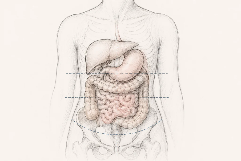
At its core, visceroptosis is a structural anomaly. The term is derived from the Latin viscera (internal organs) and the Greek ptosis (a falling or drooping). Anatomically, it refers to the condition where one or more abdominal or pelvic organs descend significantly below their expected physiological position.
While the human body is designed with a complex network of ligaments, fascia, and muscular support to keep organs in place, this support system is not infallible. In the context of connective tissue disorders, where the very "glue" of the body—collagen—is structurally compromised, the physical anchoring of the stomach, intestines, liver, and kidneys can become lax.
It is vital to distinguish between anatomic and clinical visceroptosis. Anatomic visceroptosis is a descriptive finding; an imaging scan may show a transverse colon sitting lower than average in an asymptomatic individual. Clinical visceroptosis, however, exists only when that displacement disrupts the organ’s function, leading to tangible symptoms like severe constipation, early satiety, or chronic pain. The diagnosis is not merely a matter of millimeters on a radiograph, but a correlation between structural position and physiological impairment.
A Chronological Perspective: From "Glénard’s Disease" to Obscurity
The clinical history of visceroptosis is a study in shifting medical paradigms.
The Era of Recognition (1885–1930)
In 1885, the French physician Dr. Frantz Glénard published seminal observations describing patients with "enteroptosis," a specific form of visceroptosis. His patients presented with a cluster of symptoms that were later dubbed "Glénard’s Disease." During this period, the medical community took the condition seriously. It was not uncommon for surgeons to attempt "gastropexies" or other anchoring procedures to stabilize organs. Leading journals such as The New England Journal of Medicine, JAMA, and The Lancet frequently published articles debating the etiology and management of these prolapsed organs.
The Great Disappearance
Mid-20th-century medicine pivoted toward infectious diseases, trauma, and, eventually, the rise of specialized pharmacology. The structural, mechanical view of the abdomen fell out of favor. Perhaps because the treatments of the era—which were often invasive and sometimes ineffective—could not produce consistent results, the diagnosis simply faded from textbooks. It was often dismissed as "neurasthenia," a catch-all term for patients who complained of physical symptoms that clinicians could not easily visualize or "cure" with then-current technologies.
The Modern Resurgence
In the last two decades, the conversation has been reignited, primarily through the lens of Ehlers-Danlos syndrome (EDS). Patients with hEDS often experience a cascade of systemic issues, including autonomic dysfunction and musculoskeletal pain. As these patients sought answers for their chronic gastrointestinal distress, clinicians began to revisit the concept of visceroptosis. What was once considered a psychological manifestation is now being recognized as a potential structural consequence of systemic collagen deficiency.
Supporting Data and the hEDS Connection
The intersection of hEDS and visceroptosis is perhaps the most compelling area of current research. While pelvic organ prolapse and rectal prolapse are already officially recognized within the 2017 diagnostic criteria for hEDS, the "higher" abdominal organs remain the frontier of this investigation.
Anatomical Laxity and Functional Impairment
Data suggests that the connective tissue laxity inherent in hEDS does not stop at the pelvic floor. A 2023 study published in Frontiers in Medicine involving 139 patients with slow-transit constipation revealed a statistically significant correlation between transverse colon ptosis and the severity of symptoms. While this study did not exclusively target an EDS population, it provides a crucial framework: organ position does impact motility.
In patients with hEDS, the gastrointestinal tract is already prone to dysmotility, gastroparesis, and autonomic dysfunction. When you add the mechanical burden of a displaced organ—such as a stomach that has lost its normal suspension—the result is often a compounding of symptoms. The "kinking" of a displaced colon or the physical drag of a ptotic stomach can exacerbate pre-existing transit issues, creating a cycle of pain and obstruction that is difficult to treat with standard laxatives or dietary changes alone.
The Challenge of Diagnosis
Why is this still so hard to diagnose? The answer lies in the limitations of current diagnostic protocols. Standard imaging (like a CT scan) is usually performed while the patient is lying flat (supine). Gravity, however, is the primary driver of visceroptosis. If a patient is lying down, the organs shift back into a position that may look "normal." To properly assess for visceroptosis, imaging may need to be performed in an upright or functional position—a protocol that is not standard in most radiology departments.
Implications for Patients and Clinical Practice
The implications of this "forgotten" diagnosis are profound for those living with chronic, unexplained abdominal pain.
The Burden of Dismissal
For many, the path to a diagnosis of visceroptosis is littered with years of being told their symptoms are "all in their head" or purely the result of anxiety. Because patients with hEDS are frequently dismissed as having "somatic" symptoms, the physical reality of a displaced organ is often overlooked. This pattern of medical gaslighting persists even though the mechanisms for structural laxity are well-documented in other parts of the body.
Treatment: The Cautionary Path
The approach to treatment must be conservative and highly individualized.
- Physical Therapy: Pelvic floor physical therapy and core stabilization exercises are the gold standard. Strengthening the abdominal wall can sometimes provide the "internal corset" needed to support displaced organs.
- Compression: Some patients report significant relief from abdominal compression garments, which help stabilize the abdominal wall and reduce the downward drag on the viscera.
- Surgery: This remains the most contentious area. Because patients with hEDS often have fragile tissues, surgical interventions for visceroptosis carry higher risks of poor wound healing or recurrent prolapse. Surgery should only be considered when conservative measures fail, and when the link between the ptosis and the clinical symptoms is clearly established by a specialist familiar with both EDS and structural abdominal anatomy.
Looking Forward: A Call for Evidence
The medical community is at a crossroads regarding visceroptosis. The historical records provide a foundation, but the modern era demands rigorous evidence. We need:
- Standardized Imaging: The adoption of standing or upright imaging protocols to accurately diagnose organ displacement.
- Multidisciplinary Care: Gastroenterologists, surgeons, and physical therapists must collaborate to treat the patient as a whole, rather than treating the organ in isolation.
- Further Research: Large-scale studies focusing on the prevalence of visceroptosis in the EDS population are essential to move this from anecdotal case reports to evidence-based clinical guidelines.
As we continue to learn more about the complexities of connective tissue disorders, we must ensure that the "condition medicine forgot" is finally given the attention it deserves. For the patient struggling with chronic abdominal pain, the answer may not be a new medication, but a better understanding of how their own body’s structure is interacting with the forces of gravity. By validating these experiences and refining our diagnostic approach, we can move closer to providing real, tangible relief for those who have spent too long searching for answers in the dark.
Key Takeaways
- Historical Context: Visceroptosis was a well-recognized diagnosis in the early 20th century before falling into clinical obscurity.
- The hEDS Link: Patients with connective tissue disorders are at a higher risk for organ displacement, which may contribute to chronic gastrointestinal symptoms.
- Functional vs. Anatomic: A diagnosis requires that the organ displacement actually causes functional impairment; "dropped" organs are only a problem if they cause symptoms.
- Diagnostic Gap: Standard imaging (supine) often fails to capture the true extent of organ displacement; upright imaging may be necessary.
- Treatment Caution: While conservative measures like physical therapy and compression are promising, surgery remains a complex and poorly studied option for hEDS patients due to tissue fragility.