








In a significant stride toward transforming how we diagnose and treat neurodegenerative disease, scientists at Nanyang Technological University, Singapore (NTU Singapore) have unveiled a breakthrough discovery: the brain’s primary waste disposal system often becomes obstructed in individuals showing the earliest, most subtle signs of Alzheimer’s disease.
This finding, published by the Lee Kong Chian School of Medicine (LKCMedicine), suggests that these blockages—scientifically referred to as "enlarged perivascular spaces"—act as a biological early-warning system. Because these anomalies are readily visible on routine magnetic resonance imaging (MRI) scans, the researchers believe this discovery could allow for earlier clinical intervention, potentially years before the onset of debilitating dementia symptoms.
Main Facts: The Anatomy of a Clogged Mind
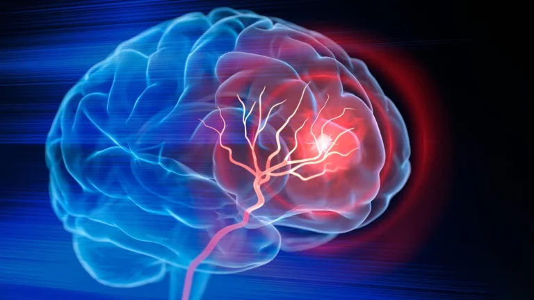
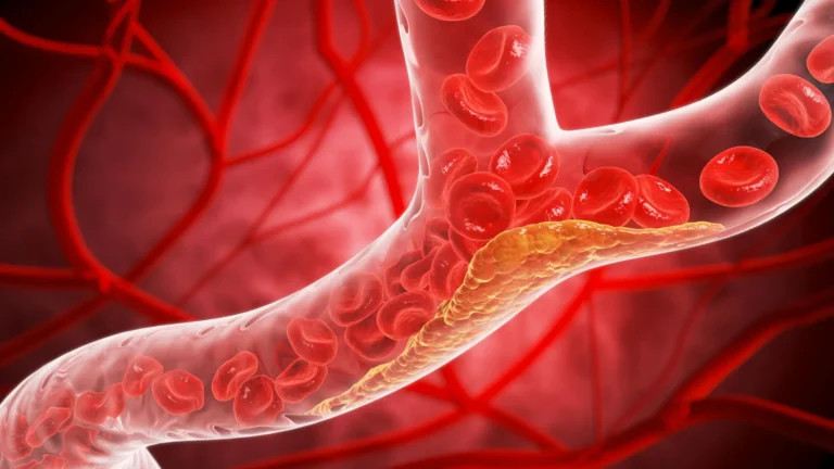
The human brain is an energy-intensive organ, and like any high-performance system, it produces metabolic waste. This waste—primarily in the form of beta-amyloid and tau proteins—is typically cleared through a network of small, fluid-filled channels known as perivascular spaces that surround the brain’s blood vessels.
When these channels function correctly, they act as a "sewage system," flushing out toxic proteins that would otherwise aggregate into the plaques and tangles associated with Alzheimer’s. However, the NTU study demonstrates that in patients with mild cognitive impairment (MCI), these spaces often become enlarged and clogged. These structural changes are not merely incidental; they are a sign that the brain’s clearance mechanism has faltered, leading to a toxic buildup that accelerates cognitive decline.
Crucially, the research highlights that these "clogs" can be identified using the same MRI scans that patients currently undergo to investigate memory loss or confusion. By shifting the clinical focus to these specific indicators, doctors may gain a diagnostic tool that is both cost-effective and readily available, avoiding the need for expensive or invasive secondary tests in the initial screening phase.
The Chronology of Discovery
The research journey was spearheaded by Associate Professor Nagaendran Kandiah, a leading authority on dementia and the Director of the Dementia Research Centre (Singapore) at LKCMedicine. The project was executed as a collaborative effort involving the school’s Bachelor of Medicine and Bachelor of Surgery program, with fifth-year student Justin Ong serving as the study’s first author.
Phase 1: Population-Specific Analysis
The team recognized a significant gap in global Alzheimer’s research: the overwhelming reliance on Caucasian cohorts. Recognizing that dementia manifests differently across ethnic groups, the researchers recruited nearly 1,000 participants from Singapore’s diverse demographic. This was a deliberate attempt to move away from Western-centric data, which often fails to account for regional genetic variations, such as the prevalence of the apolipoprotein E4 gene.
Phase 2: Diagnostic Comparison
The study bifurcated participants into two primary groups: those with normal cognitive function (the control group) and those exhibiting mild cognitive impairment. Researchers performed high-resolution MRI scans to map the perivascular spaces and cross-referenced these findings with blood-based biochemical markers for Alzheimer’s—specifically measuring levels of seven proteins known to be pathological indicators.
Phase 3: The "Smoking Gun"
As the team analyzed the data, they discovered that the presence of enlarged perivascular spaces correlated strongly with four of the seven blood-based markers of Alzheimer’s. Even more tellingly, when compared against the "gold standard" of white matter damage (a common indicator of vascular health), the perivascular blockages emerged as a more sensitive predictor of Alzheimer’s-related pathology in the early stages of cognitive decline.
Supporting Data: Why Regional Research Matters
The necessity for this study arose from a fundamental biological discrepancy identified by Prof. Kandiah. In Caucasian populations, the apolipoprotein E4 gene—a major genetic risk factor for Alzheimer’s—is found in approximately 50 to 60 percent of patients with the disease. In the Singaporean cohort studied by the NTU team, that figure plummeted to less than 20 percent.
This discrepancy serves as a stark reminder that Alzheimer’s is not a monolithic disease. By focusing on an Asian population, the researchers have provided data that is not only more accurate for local medical practice but also highlights the nuance required in global neurodegenerative research.
The data further revealed that the correlation between perivascular space enlargement and blood-based biomarkers was particularly potent in patients with MCI. This suggests that the "clogging" process is a primary driver of disease progression, rather than a late-stage byproduct. The team found that as these spaces enlarged, the systemic level of toxic proteins rose, indicating a direct, synergistic relationship between vascular health and neurological integrity.
Official Responses and Clinical Perspectives
The scientific community has received the study with cautious optimism, noting that it provides a much-needed bridge between vascular medicine and neurology.
Dr. Rachel Cheong Chin Yee, Senior Consultant and Deputy Head at the Department of Geriatric Medicine at Khoo Teck Puat Hospital, emphasized the clinical utility of the findings. "These findings are significant because they suggest that brain scans showing enlarged perivascular spaces could potentially help identify people at higher risk of Alzheimer’s disease, even before symptoms appear," Dr. Cheong stated. She noted that this adds a layer of predictive power to routine diagnostics.
Dr. Chong Yao Feng, a Consultant at the National University Hospital’s Division of Neurology, offered a perspective on the integration of these findings into standard practice. "The study’s findings are intriguing as they demonstrate that both cerebrovascular diseases and Alzheimer’s interact in a synergistic manner," said Dr. Chong. He cautioned, however, that physicians must exercise clinical judgment. The presence of these markers should trigger a conversation between doctor and patient, leading to a more nuanced diagnostic pathway rather than an automatic diagnosis.
Implications: A New Era for Early Intervention
The implications of this research are twofold: diagnostic efficiency and the timing of medical intervention.
1. Refined Diagnostic Pathways
Currently, clinicians often look for "white matter hyperintensities"—scars or damage in the brain’s white matter—when interpreting MRI scans for signs of dementia. While useful, this approach can sometimes overlook the subtle early signs of Alzheimer’s. By adding the assessment of perivascular spaces to the standard radiology checklist, doctors can capture "at-risk" patients much earlier.
2. A Window of Opportunity
"Identifying Alzheimer’s sooner gives doctors more time to intervene and potentially slow the progression of symptoms such as memory loss, reduced thinking speed, and mood changes," says Justin Ong. The window between the first signs of cognitive impairment and the onset of clinical dementia is the most critical time for therapeutic intervention. By identifying the physical obstruction of waste removal, physicians may eventually be able to prescribe treatments—ranging from lifestyle modifications to emerging pharmacological agents—that target the root causes of this clogging.
Future Directions: Tracking the Trajectory
The work of the NTU team is far from finished. The researchers have committed to a long-term follow-up study, tracking the participants over the coming years. By observing how many individuals with enlarged perivascular spaces eventually transition from mild cognitive impairment to full-blown Alzheimer’s dementia, the team hopes to validate these scans as a definitive prognostic tool.
If these longitudinal results confirm the initial findings, the diagnostic landscape for Alzheimer’s could change permanently. A routine MRI, once considered a tool primarily for ruling out tumors or strokes, may soon become the definitive instrument for assessing the "drainage health" of the human brain.
As the global population ages, the burden of Alzheimer’s disease continues to rise, placing an unprecedented strain on healthcare systems. The NTU Singapore study provides a flicker of hope—a way to see the disease coming before it arrives, allowing for the potential to preserve cognitive function and improve the quality of life for millions of people worldwide. The "clogged drain" of the brain may finally have a solution, provided we are willing to look closely enough at the images already on our screens.