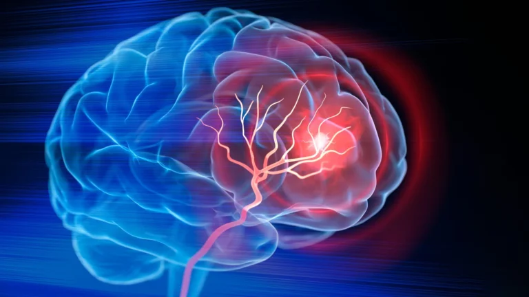










In the silent, intricate architecture of the human brain, a sophisticated waste-removal system works tirelessly to clear out toxic proteins that threaten cognitive longevity. New research from Nanyang Technological University, Singapore (NTU Singapore) has unveiled a critical vulnerability in this system: when these drainage pathways become clogged, it may serve as an early, visual warning signal for Alzheimer’s disease, often years before debilitating symptoms manifest.
This discovery offers a potential paradigm shift in neurology. By identifying "enlarged perivascular spaces" on routine brain scans, clinicians may soon be able to flag patients at risk of cognitive decline long before the onset of clinical dementia, providing a crucial window for medical intervention.
The Mechanism of Failure: Clogged Brain Drains
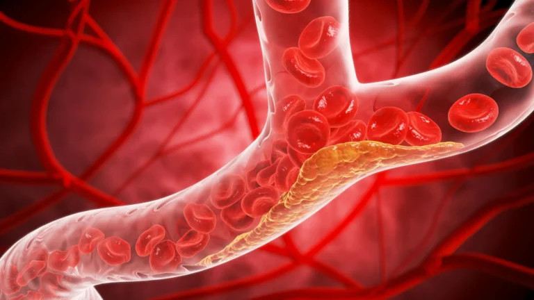
Deep within the brain, blood vessels are enveloped by microscopic channels known as perivascular spaces. These spaces function as a vital drainage network, flushing out neurotoxic metabolic byproducts—most notably beta-amyloid and tau proteins—which are hallmark drivers of Alzheimer’s disease.
Under healthy conditions, these channels are fluid and efficient. However, the NTU study indicates that in the early stages of cognitive decline, these pathways frequently become blocked or enlarged. When the brain’s “plumbing” fails, these toxic proteins accumulate, setting the stage for neurodegeneration.
Until now, the clinical significance of these enlarged spaces remained a subject of academic debate. While they were known to appear on magnetic resonance imaging (MRI) scans, it was unclear whether they were a direct indicator of Alzheimer’s pathology or merely a byproduct of general aging. The NTU team, led by Associate Professor Nagaendran Kandiah of the Lee Kong Chian School of Medicine (LKCMedicine), has effectively bridged this gap by correlating these visual anomalies with established biochemical markers of the disease.
A Chronology of Discovery: From Observation to Clinical Insight
The path to this discovery was rooted in a comprehensive longitudinal effort to understand cognitive decline within diverse Asian populations. The research, which involved nearly 1,000 participants from various ethnic backgrounds in Singapore, utilized a robust methodology to ensure the results were both accurate and representative.
The Research Framework
The study was conducted as part of the LKCMedicine’s Scholarly Project module, led by fifth-year medical student and primary author Justin Ong. The researchers segmented the cohort into two distinct groups: those with normal cognitive function and those exhibiting early signs of mild cognitive impairment (MCI).
- Initial Screening: The team performed standard MRI scans on all participants to identify the presence of enlarged perivascular spaces.
- Biochemical Profiling: To validate the MRI findings, the team measured seven key blood-based biochemical markers known to be associated with Alzheimer’s, including elevated levels of beta-amyloid and tau proteins.
- Comparative Analysis: The researchers compared the prevalence of enlarged perivascular spaces against traditional indicators of brain health, such as white matter damage—the degradation of nerve fiber connections that typically signal vascular issues in the brain.
The data revealed a startling correlation: individuals with mild cognitive impairment were significantly more likely to display these enlarged spaces than their healthy counterparts. Furthermore, these "clogs" showed a stronger statistical link to Alzheimer’s-related blood markers than the traditional white matter damage markers, suggesting that these spaces are a more sensitive, early-stage indicator of the disease.
The Vital Importance of Asian-Centric Research
A significant portion of the global scientific discourse on Alzheimer’s has historically relied on data derived from Caucasian populations. However, the NTU study underscores why this "one-size-fits-all" approach is fundamentally flawed.
Assoc Prof Kandiah, who also serves as the Director of the Dementia Research Centre (Singapore), points to the genetic disparities that necessitate region-specific research. For instance, the apolipoprotein E4 (APOE4) gene, a major risk factor for Alzheimer’s, is present in 50 to 60 percent of Caucasian dementia patients. In contrast, that same gene is found in less than 20 percent of Singaporean patients.
"Because of these differences, findings in one population may not directly apply to another," Kandiah notes. By focusing on a multi-ethnic Asian cohort, the NTU study provides a more nuanced understanding of how Alzheimer’s manifests across different genetic and environmental backdrops, making the findings far more applicable to the global population than previous Western-centric models.
Implications for Clinical Practice and Early Intervention
The diagnostic implications of this study are profound. Currently, detecting Alzheimer’s often requires expensive, invasive, or time-consuming tests, such as positron emission tomography (PET) scans or lumbar punctures to measure cerebrospinal fluid.
A Cost-Effective Diagnostic Tool
Because enlarged perivascular spaces are visible on standard MRI scans, their use as a biomarker could revolutionize clinical practice. "Identifying them could complement existing methods to detect Alzheimer’s earlier, without having to do and pay for additional tests," explains Assoc Prof Kandiah.
The Window for Intervention
Early detection is the cornerstone of modern geriatric medicine. Justin Ong emphasizes that spotting these signs early provides doctors with the critical time needed to implement lifestyle changes, pharmacological treatments, or dietary adjustments that could potentially slow the progression of memory loss, executive dysfunction, and mood changes.
Official Responses and Expert Perspective
The medical community has reacted to these findings with a mix of optimism and cautious interest, noting that the study challenges the traditional separation between vascular disease and neurodegenerative disorders.
Dr. Rachel Cheong Chin Yee, a Senior Consultant and Deputy Head at Khoo Teck Puat Hospital’s Department of Geriatric Medicine, suggests that this research forces a re-evaluation of how we categorize "small vessel disease" in the brain. "These findings are significant because they suggest that brain scans showing enlarged perivascular spaces could potentially help identify people at higher risk of Alzheimer’s disease, even before symptoms appear," Dr. Cheong stated.
Similarly, Dr. Chong Yao Feng, a Consultant at the National University Hospital’s Division of Neurology, notes that the study highlights a "synergistic" relationship between cerebrovascular health and Alzheimer’s pathology. Traditionally, doctors might have attributed cognitive decline solely to vascular issues if they saw signs of vessel damage on a scan. Dr. Chong cautions that clinicians must now look deeper.
"Doctors will then have to use their clinical judgement of the patient’s scan and symptoms," Dr. Chong advised. "They must discuss with the patient to determine if more checks are needed to confirm whether a patient has Alzheimer’s disease or not, rather than assuming it is purely a vascular issue."
The Road Ahead: Future Research and Validation
While the results are compelling, the research team is not stopping here. The next phase of the study involves long-term tracking of the participants to observe how many eventually transition from mild cognitive impairment to full-blown Alzheimer’s dementia. This longitudinal data will be essential in confirming the predictive power of the enlarged perivascular spaces.
If these findings are replicated in further studies across other global populations, the routine examination of perivascular spaces on MRI scans could become a standard practice in geriatric screenings worldwide. This could transform the Alzheimer’s patient journey from one of late-stage diagnosis and symptom management to one of proactive, early-stage intervention.
By turning the lens toward the brain’s own waste-disposal system, the NTU Singapore researchers have provided the medical community with a new, accessible, and potentially life-saving tool in the fight against one of the most challenging health crises of the 21st century. The path toward a world where Alzheimer’s can be caught before it steals a person’s memories is becoming clearer, one scan at a time.