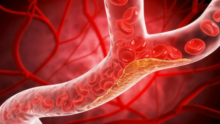


For decades, the standard laboratory model for studying the human circulatory system has relied on a fundamental—and often misleading—simplification: the straight, uniform tube. While these models provided a baseline for understanding basic fluid dynamics, they failed to account for the chaotic, intricate architecture of the human vascular system. In reality, blood vessels are dynamic conduits that branch, twist, narrow, and expand. These architectural complexities are not merely incidental; they are the primary sites where vascular diseases, such as aneurysms and stenoses, take root.
Now, a team of researchers at the Texas A&M University Department of Biomedical Engineering is challenging these limitations. By developing a highly customizable, "living" vessel-chip system, the Bioinspired Translational Microsystems Laboratory is bridging the gap between simplified lab models and the complex reality of human physiology. This breakthrough, recently published in the journal Lab on a Chip and slated for the May 2025 cover, offers a powerful new platform for drug testing, disease modeling, and personalized medicine.
The Architectural Gap: Why Geometry Matters in Vascular Health
The human body’s circulatory system is a masterpiece of fluid engineering, yet it is highly susceptible to structural degradation. When a blood vessel branches, the flow of blood is disrupted, creating zones of turbulent or recirculating flow. Similarly, when a vessel narrows (stenosis) or balloons out (aneurysm), the physical forces acting upon the vessel walls change dramatically.
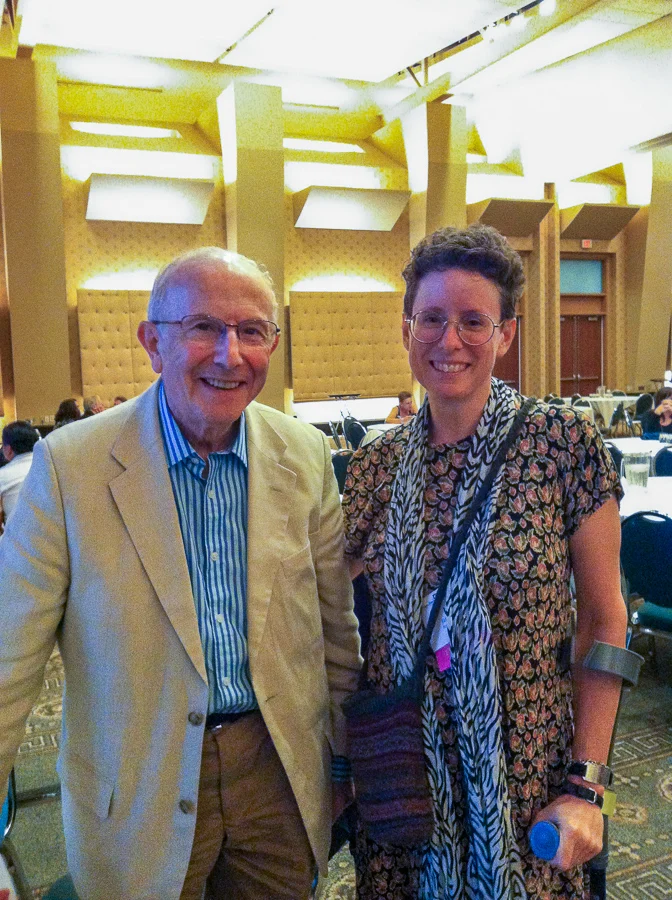
"There are branched vessels, or aneurysms that have sudden expansion, and then stenosis that restricts the vessel," explains Jennifer Lee, a master’s student in biomedical engineering and the lead researcher on the project. "All these different types of vessels cause the blood flow pattern to be significantly changed, and the inside of the blood vessel is affected by the level of shear stress caused by these flow patterns. That’s what we wanted to model."
Shear stress—the frictional force exerted by blood flow against the endothelial lining—is a critical factor in maintaining vascular health. When shear stress becomes irregular due to complex geometry, it can trigger inflammatory responses, plaque buildup, and other pathological conditions. Traditional straight-tube models simply cannot replicate these nuanced biomechanical environments. By moving beyond the straight line, the Texas A&M team is finally creating an environment where these localized disease triggers can be studied in a controlled, repeatable manner.
A Chronology of Innovation: From Basic Models to Bio-Complexity
The journey to the current vessel-chip iteration has been one of incremental, yet transformative, scientific progress.
The Foundation (2020–2022)
The project traces its lineage to earlier work conducted in Dr. Abhishek Jain’s Bioinspired Translational Microsystems Laboratory. A few years prior, Dr. Tanmay Mathur, then a graduate student, successfully developed a straight vessel-chip design. This early model was an essential proof-of-concept, demonstrating that microfluidic devices could effectively mimic the basic interactions between endothelial cells and blood flow. It provided the framework for the lab’s core competency in organ-on-a-chip technology.
The Developmental Phase (2023–2024)
Recognizing the limitations of the straight-tube design, Jennifer Lee took up the mantle of advancing the platform. Joining the lab as an undergraduate honors student, Lee was tasked with creating a system that could replicate the wide range of structural geometries seen in the human body. Her work involved rigorous testing of materials and micro-engineering techniques to ensure the chips could withstand the fluid pressures required to mimic realistic blood flow while maintaining the viability of living tissue.
The Breakthrough (2025)
The resulting platform is a modular, customizable system that can be tailored to replicate specific patient anatomies. The research, detailing the methodology for creating these complex micro-architectures, was accepted for publication in Lab on a Chip. The upcoming May 2025 cover feature serves as a testament to the scientific community’s recognition of the platform’s significance in the field of vascular biology.
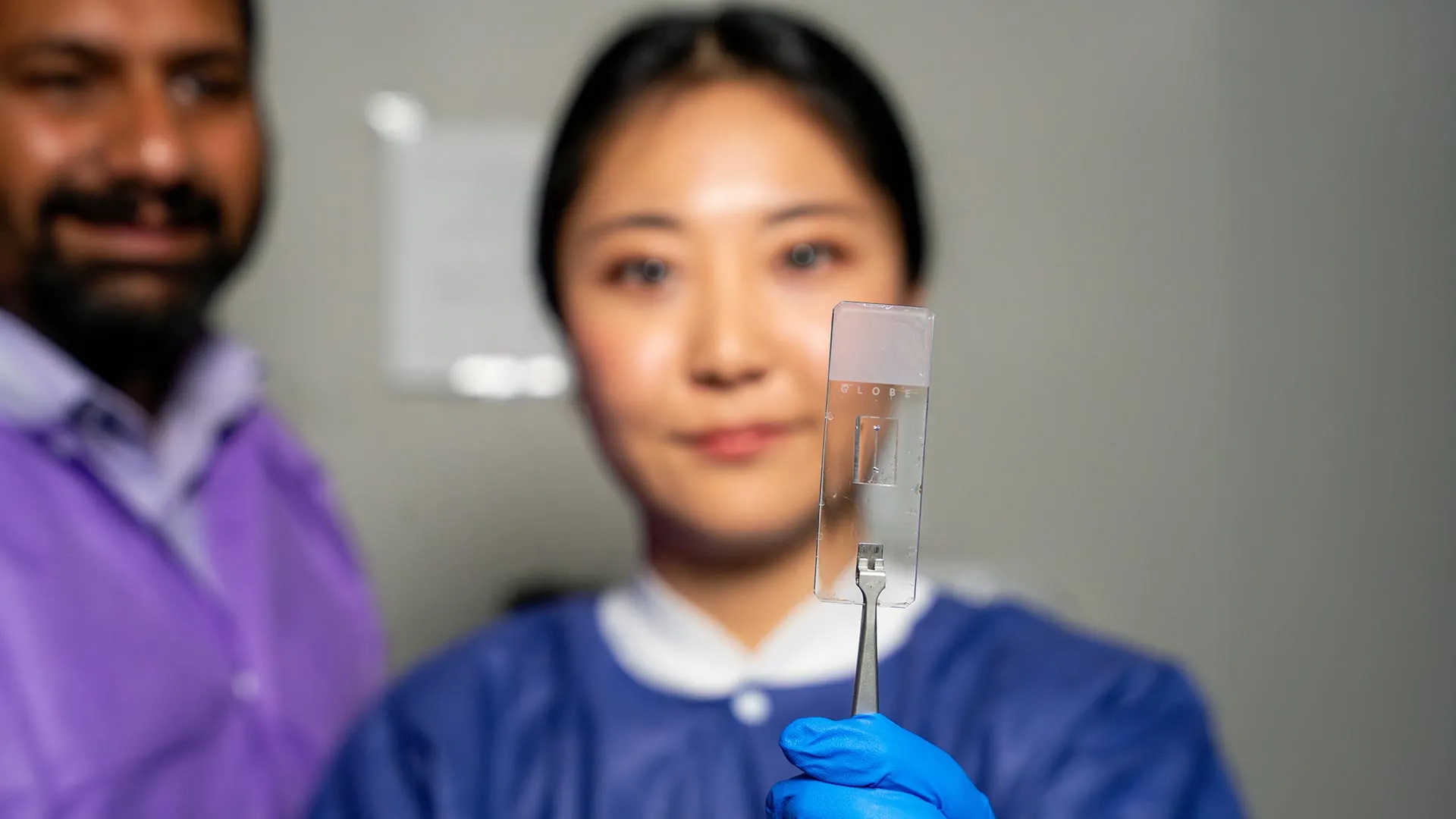
Supporting Data and Technical Significance
The vessel-chip technology functions by using microfluidics to create microscopic channels lined with living human endothelial cells. These devices are more than just plastic molds; they are "living" systems.
Key features of the current platform include:
- Geometric Customization: Unlike static, pre-cast models, the Texas A&M device can be engineered to mimic specific pathological states, including bifurcations (branching), stenotic narrowing, and aneurysmal dilation.
- Endothelial Integration: The chips are lined with functional endothelial cells, the critical barrier between blood and the vessel wall. This allows for real-time observation of how these cells respond to varying levels of shear stress.
- High-Fidelity Flow Dynamics: The system allows researchers to precisely control the velocity and pattern of fluid flow, replicating both healthy physiological states and diseased, turbulent states.
The ability to incorporate actual human cellular material is what elevates this from a mechanical simulation to a biological tool. As Dr. Abhishek Jain, associate professor and the Barbara and Ralph Cox ’53 faculty fellow, notes: "Not only can you make these structures complex, you can put actual cellular and tissue material inside them and make them living. These are the sites where vascular diseases tend to develop, so understanding them is critical."
Official Perspectives: The Vision for the Future
The success of the vessel-chip project is viewed by the Texas A&M faculty as a major milestone for the university’s fast-track research programs. The initiative is designed to push students beyond the scope of traditional undergraduate projects, encouraging them to pursue high-impact, high-risk research with tangible outcomes.
"Jennifer demonstrated perseverance, curiosity, and creativity and started taking up research projects very quickly," said Dr. Jain. "Our fast-track program enables students like Jennifer to take on sort of high-impact, high-risk research and not just do a science project, but take it all the way to its outcome and get it published."
Looking toward the horizon, the research team is already conceptualizing the "fourth dimension" of organ-on-a-chip technology. While the current iteration focuses on endothelial cells, future versions aim to incorporate multi-cellular layers, including smooth muscle cells and immune components. This would allow for a more comprehensive study of how different tissue types interact with each other and with blood flow under pathological stress.
"We are progressing and creating what we call the fourth dimensionality of organs-on-a-chip, where we not only focus on the cells and the flow, but this interaction of cells and flow in more complex architectural states, which is a new direction in the field," Dr. Jain added.
Implications for Modern Medicine
The implications of this technology are vast, particularly in the realm of personalized medicine and pharmacological development.
Reducing Animal Dependency
One of the most significant advantages of vessel-chips is their potential to reduce the reliance on animal models. Animal biology often fails to perfectly mirror human vascular responses to drugs; by using human-derived cells in a chip that mimics human architecture, researchers can achieve higher predictive accuracy for how a new treatment will perform in a clinical setting.
Accelerating Drug Discovery
Testing new drugs on "living" complex vessels allows pharmaceutical researchers to observe side effects or efficacy issues in a controlled environment before moving to human trials. This could significantly reduce the cost and time associated with the drug development pipeline.
Personalized Disease Modeling
Because these chips can be tailored to individual patient anatomies—potentially using cells derived from the patient themselves—they open the door to "precision vascular medicine." Doctors could theoretically test how a patient’s specific vessel structure responds to a variety of medications, allowing for the selection of the most effective therapy with the fewest side effects.
The Human Element: Training the Next Generation
Beyond the technical breakthroughs, the vessel-chip project highlights the importance of institutional research environments. For Jennifer Lee, the experience was about more than just mastering microfluidics; it was an exercise in professional development.
The lab’s collaborative atmosphere, involving postdoctoral researchers, graduate students, and peers, provided a space to refine skills in communication, teamwork, and ethical research practices. "It’s such a good environment to interact with not only peers but also graduate students and postdoctoral researchers," Lee said. "You’re able to learn teamwork and communication, work ethic, and just trying different things out. I think it’s such a valuable experience that students have available."
This holistic approach to education ensures that as the technology moves from the lab bench to potential clinical application, the scientists behind it are equipped to lead, collaborate, and innovate in a global scientific community.
Conclusion: A New Standard for Vascular Research
The work emerging from Texas A&M’s Bioinspired Translational Microsystems Laboratory represents a paradigm shift in how we understand the circulatory system. By embracing the complexity of human biology rather than ignoring it, researchers have developed a tool that is not only scientifically rigorous but also profoundly practical.
With support from a coalition of federal and scientific entities—including the U.S. Army Medical Research Program, NASA, the NIH, the FDA, and the National Science Foundation—the vessel-chip is poised to become a staple of vascular research. As the team continues to refine their "living" models, the gap between the laboratory and the clinic continues to shrink, promising a future where vascular disease is better understood, better treated, and more effectively prevented.